The [KO Validated] CDKN1A/p21CIP1 Antibody (CAB2691) is a high-quality antibody developed for reliable detection and analysis of target proteins. This antibody, produced using rabbit immunization, has high specificity for human samples and has been validated for use in Western blot assays. By targeting the CDKN1A protein, this antibody allows for precise detection and analysis of protein expression levels in a variety of cell types, making it ideal for investigations in cell biology and cancer research.
This antibody is validated for use in WB, IHC-P, IP, ELISA applications and has demonstrated reactivity against Human, Mouse samples.
Product Name:
[KO Validated] CDKN1A/p21CIP1 Antibody
SKU:
CAB2691
Size:
20μL, 100μL
Reactivity:
Human, Mouse
Conjugate:
Unconjugated
Immunogen:
Recombinant protein (or fragment).This information is considered to be commercially sensitive.
This gene encodes a potent cyclin-dependent kinase inhibitor. The encoded protein binds to and inhibits the activity of cyclin-cyclin-dependent kinase2 or -cyclin-dependent kinase4 complexes, and thus functions as a regulator of cell cycle progression at G1. The expression of this gene is tightly controlled by the tumor suppressor protein p53, through which this protein mediates the p53-dependent cell cycle G1 phase arrest in response to a variety of stress stimuli. This protein can interact with proliferating cell nuclear antigen, a DNA polymerase accessory factor, and plays a regulatory role in S phase DNA replication and DNA damage repair. This protein was reported to be specifically cleaved by CASP3-like caspases, which thus leads to a dramatic activation of cyclin-dependent kinase2, and may be instrumental in the execution of apoptosis following caspase activation. Mice that lack this gene have the ability to regenerate damaged or missing tissue. Multiple alternatively spliced variants have been found for this gene.
Purification Method
Affinity purification
Gene ID
1026
RRID
AB_2863018
Buffer Information
Store at -20℃. Avoid freeze / thaw cycles. Buffer: PBS with 0.09% Sodium azide,50% glycerol,pH7.3.
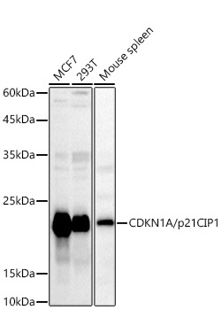
Western blot analysis of various lysates using CDKN1A/p21CIP1 Rabbit pAb (CAB2691) at 1:1000 dilution. Secondary antibody: HRP-conjugated Goat anti-Rabbit IgG (H+L) (CABS014) at 1:10000 dilution. Lysates/proteins: 25μg per lane. Blocking buffer: 3% nonfat dry milk in TBST. Detection: ECL Basic Kit (AbGn00020). Exposure time: 180s.
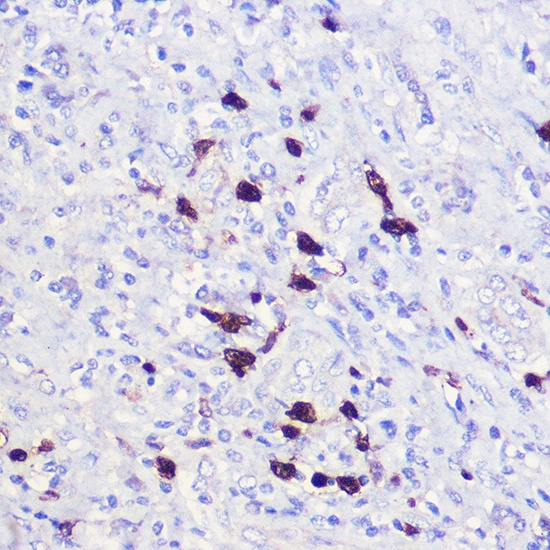
Immunohistochemistry analysis of paraffin-embedded Human liver using CDKN1A/p21CIP1 Rabbit pAb (CAB2691) at dilution of 1:100 (40x lens). High pressure antigen retrieval performed with 0.01M Citrate buffer (pH 6.0) prior to IHC staining.
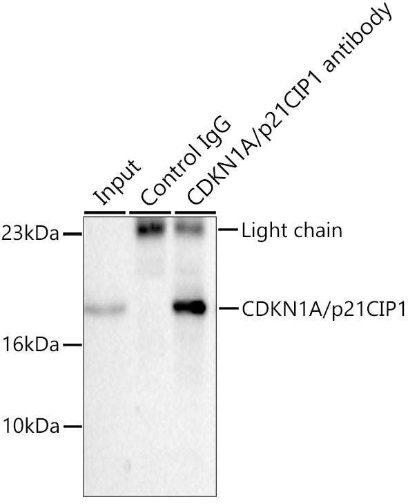
Immunoprecipitation of CDKN1A/p21CIP1 from 200 µg extracts of MCF7 cells was performed using 0.5 µg of CDKN1A/p21CIP1 Rabbit pAb (CAB2691). Rabbit IgG isotype control (AC042) was used to precipitate the Control IgG sample. IP samples were eluted with 1X Laemmli Buffer. The Input lane represents 10% of the total input. Western blot analysis of immunoprecipitates was conducted using CDKN1A/p21CIP1 Rabbit pAb (CAB2691) at a dilution of 1:500.