 Assay Kit (BR00001)")
Description
Fatty Acid Oxidation Assay Kit
The Assay Genie Fatty Acid Oxidation Assay kit is a highly sensitive colorimetric assay for the quantitative measurement of Fatty Acid Oxidation in cells and tissues. This unique assay utilizes a highly soluble Octanoyl-CoA substrate which enables the accurate measurement of Fatty Acid Oxidation versus longer chain less-soluble substrates such as the 18C unsaturated fatty acid oleate.
| Product Name: | Fatty Acid Oxidation Assay |
| SKU: | BR00001 |
| Application: | Quantitative measurement of fatty acid oxidation in cells and tissues. |
| Note: | For Research Use Only |
- Fatty acids provide more ATP per carbon than carbohydrates such as glucose. In tissues with high energy requirements such as heart tissue, up to 50 – 70% of energy comes from fatty acid beta-oxidation, which converts fatty acids to acetyl-CoA with concomitant production of FADH2 and NADH in the mitochondria.
- This strictly aerobic oxidative pathway consists of four distinct steps:
- Acyl-CoA dehydrogenation by acyl-CoA dehydrogenase
- Hydration by enoyl-CoA hydratase
- Dehydrogenation by 3-hydroxyacyl-CoA dehydrogenase
- Thiolytic cleavage by beta-ketothiolase
- The importance of the beta-oxidation system in humans is exemplified by the existence of a group of genetic diseases categorized as fatty acid oxidation disorders (FAODs), which are a heterogeneous group of defects in fatty acid transport and oxidation, and are inherited as autosomal recessive disorders, having a wide range of clinical presentations.
- Evidence indicates that regulation of activity of FAO during fetal development is not only important for fetal life but also has implications for health and disease in adulthood.
- The measurement of FAO activity provides valuable insights into the pathophysiologic and metabolic basis of many diseases.
- The Assay Genie non-radioactive FAO assay is based on oxidation of the substrate octanoyl-CoA. Generation of NADH is coupled to the reduction of the tetrazolium salt INT to formazan. The intensity of the red-colored formazan is proportional to increased FAO activity.
- The assay solution and substrate should be stored in aliquots at -80ºC.
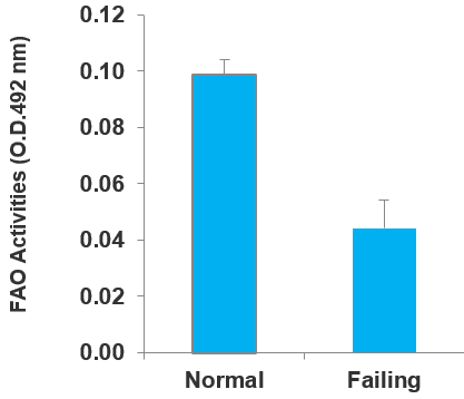
Figure 1: Tissue lysates were prepared from healthy Hamster hearts and the hearts of the TO2 Hamster model which is associated with heart failure. Lysates were used in the assay and the levels of fatty acid oxidation quantified.
Figure 2: Octanoyl-CoA is the substrate, not the product of the reaction. Please see the diagram.
Preparation of cell/tissue extracts:
| Step | Procedure |
| Step 1. | Wash ~106 cells twice with ice-cold phosphate-buffered saline (PBS), and remove PBS completely from the cell pellet. Cell pellet should be stored at -80ºC. Tissue sample should be washed with PBS thoroughly to remove blood cells, which can cause inconsistent assay result. |
| Step 2. | Prepare enough 1x Cell Lysis Solution by diluting 10x Cell Lysis Solution with ice-cold dH2O. Add 50 – 100 µl ice-cold 1x Cell Lysis Solution to cell pellet. Extract cells by pipetting up and down (gently but thoroughly). Leave lysate on ice for 5 min with intermittent gentle agitation. If lysate is viscous, add more 1x Cell Lysis Solution and repeat pipetting. Centrifuge lysate in a refrigerated microfuge for 5 min at maximal speed. Recover supernatant for assay. Tissue is homogenized in 1x Cell Lysis Solution, and lysate is clarified by centrifugation. Use ~25 mg of tissue per 0.5 ml of 1x Cell Lysis Solution for tissue homogenization. |
| Step 3. | Perform protein assay to determine sample protein concentration. Normalize sample protein concentration by diluting with ice-cold 1x Cell Lysis Solution to 1 – 3 mg/ml. Keep protein sample on ice at all times. Freeze thawed crude protein lysate can exhibit reduced enzyme activity. Lysate should be stored at -80ºC. Note: Do not use a buffer containing reducing agents or SDS. |
Reagent thawing:
Keep thawed FAO Assay Solution and 20x FAO Substrate on ice. Gently agitate solution prior to pipetting. It is important to minimize the time the reagents are thawed. Freeze solutions immediately after use.
Preparation of control solution and reaction solution:
- Control solution is prepared by mixing 1 part of dH2O and 20 parts of FAO Assay Solution, e.g., 25 µl dH2O mixed with 500 µl FAO Assay Solution. Keep freshly prepared control solution on ice during assay.
- Reaction solution is prepared by mixing 1 part of 20x FAO substrate and 20 parts of FAO Assay Solution, e.g., 25 µl 20x FAO Substrate mixed with 500 µl FAO Assay Solution. Keep solution on ice and use immediately. Each protein sample is treated with 50 µl control solution and 50 µl reaction solution in two separate sets.
| Step | Procedure |
| Step 1. | Add 10 µl of each protein sample to a 96-well plate in duplicate. Note: For drug discovery applications, add 1 µl of a drug inhibitor to both wells, and mix with sample by pipetting up and down. |
| Step 2. | After all samples have been pipetted to the plate, swiftly add 50 µl control solution to one set of wells and 50 µl reaction solution to another set of wells. Mix contents by gentle agitation for 30 sec. Cover plate and incubate in a humidified 37°C incubator for 1 − 2 hr. FAO activity generates cherry red color in wells. Do not use a CO2 incubator. Note: Increase sample protein concentration and/or incubation time if little red color is detected. |
| Step 3. | Stop assay by adding 50 µl 3% Acetic acid (not included in the kit) to each control solution well and reaction solution well followed by brief gentle agitation. Measure O.D.492 nm using a plate reader. |
| Step 4. | Subtract control well reading from reaction well reading for each sample. The subtracted O.D. reading is proportional to fatty acid oxidation activity of the sample. |
1) Have you tested with cells? If not, how do you know 106 is the right number? Could you use less?
Since FAO activity is driven by mitochondrial contents, most cultured cells exhibit low, but detectable, FAO activity. 106 cells are required to make a concentrated protein extract for FAO detection.
2) Will the assay work with frozen tissue? Does it not depend on active enzymes for assay to work?
The assay works with frozen tissue. It is an enzyme activity assay.
3) How do you generate a standard curve to determine your results?
There is no standard curve for the assay. Optical density can be converted to enzyme units.
4) What inhibitor can you use for FAO?
We have not tested an FAO inhibitor.
5) What controls do you recommend for the assay especially with cells in culture?
Cells/tissues with high mitochondrial contents can be used as positive control.
6) Would this assay potentially work in whole blood samples or purified PBMCs?
The red colour of the sample will interfere with optical detection at 492nm. PBMC, if free of blood cells, can be processed using the standard protocol.
7) Can this measure Fatty Acid Oxidation in cells and tissues in plant material?
The assay has not been tested with plant cells, but I would expect it to work because this is a chemical rather than immunological assay.
8) Does this assay work with extracellular fluids?
The assay does not work with extracellular fluids.
9) Is measuring Long-chain FA a more accurate determination of FAO? As short and medium do not need mitochondrial CPTs, while long ones do.
No, CPT is only relevant in an intact organ/cell system wherein the mitochondrial structure is maintained. The FAO assay however is based on the use of tissue/cell homogenates, and the activity of CPT therefore cannot be measured in the in vitro assay system.
10) How does the kit distinguish NADH production from the octanoyl-CoA from NADH production from endogenous tissue substrates (glycogen, triglycerides, etc.)?
The control solution prepared according to the protocol will measure NADH production from endogenous tissue substrates. The optical density obtained with the control solution is then subtracted out in final calculation of FAO activity.
11) It doesn’t look like there are standards. The FAO activity is calculated. How do I generate a ΔOD at 30 minutes if I’m only to measure at 30 and 60? Do I measure at time 0 as well?
You can measure OD without stopping the reaction at 30, 60, 90, or 120 min.
12) The 10x cell lysis solution has a lot of white sediment on the bottom that doesn’t seem to be dissolving. Should I warm the solution to resolubilize things?
Yes, it is caused by dry ice freezing. Just warm the solution with agitation.
- A 3% Acetic acid solution needs to be prepared for reaction termination. The assay solution contains DMSO and iodonitrotetrazolium violet. Please refer to the product page of our website or contact us for MSDS information.
| Standage et al. | NMR-Based Serum and Urine Metabolomic Profile Reveals Suppression of Mitochondrial Pathways in Experimental Sepsis-Associated Acute Kidney Injury | American Journal of Physiology-Heart and Circulatory Physiology 2021 | View Citation |
| Trompette et al. | Gut-derived short-chain fatty acids modulate skin barrier integrity by promoting keratinocyte metabolism and differentiation | Mucosal Immunology 2022 | PubMed ID: 35672452 |
| Rajala et al. | Insulin-like growth factor 1 receptor mediates photoreceptor neuroprotection | Cell Death & Disease 2022 | PubMed ID: 35840554 |
| Pham et al. | Neutrophil trafficking to the site of infection requires Cpt1a-dependent fatty acid β-oxidation | Communications Biology 2022 | PubMed ID: 36513703 |
| Quentin et al. | Oleoylethanolamide improves energy disposal in a cellular model of Alzheimer’s disease | Journal of Mitochondria, Plastids and Endosymbiosis 2024 | View Citation |
| Sun et al. | ALKBH5-mediated upregulation of CPT1A promotes macrophage fatty acid metabolism and M2 macrophage polarization, facilitating malignant progression of colorectal cancer | Experimental Cell Research 2024 | PubMed ID: 38479704 |
| Fuwa et al. | Mitochondrial fractions located in the cytoplasmic and peridroplet areas of white adipocytes have distinct roles | FEBS letters 2024 | PubMed ID: 38658180 |
| El-Hachem et al. | Valine aminoacyl-tRNA synthetase promotes therapy resistance in melanoma | Molecular Oncology and Drug Resistance 2024 | PubMed ID: 38849541 |
| Xu et al. | Phytic acid-based nanomedicine against mTOR represses lipogenesis and immune response for metabolic dysfunction-associated steatohepatitis therapy | Nanomedicine and Metabolic Disease 2024 | View Citation |
| Dipasree Hajra et al. | Salmonella-induced SIRT1 and SIRT3 are crucial for maintaining the metabolic switch in bacteria and host for successful pathogenesis | Microbial Pathogenesis and Host Metabolism 2022 | View Citation |
| Firth et al. | The Role of Mitochondrial Translocator Protein 18kda (TSPO) in Regulating Astrocyte Metabolism | Neuroscience and Metabolic Regulation 2023 | View Citation |
| Peng Wang et al. | Berberine alleviates non-alcoholic hepatic steatosis partially by promoting SIRT1 deacetylation of CPT1A in mice | Gastrointestinal and Liver Metabolism 2023 | View Citation |
| Lixia Chen et al. | The lipid-metabolism enzyme ECI2 reduces neutrophil extracellular traps formation for colorectal cancer suppressio | Nature Communications 2024 | View Citation |
| Peihao Liu et al. | Ubiquitin-specific peptidase 25 ameliorates hepatic steatosis by stabilizing peroxisome proliferator activated receptor alpha | Journal of Biological Chemistry 2024 | View Citation |
| Duraipandy Natarajan et al. | Chronic β3-AR stimulation activates distinct thermogenic mechanisms in brown and white adipose tissue and improves systemic metabolism in aged mice | Aging Cell 2024 | View Citation |
| Duraipandy Natarajan et al. | The metabolic benefits of thermogenic stimulation are preserved in aging | bioRxiv 2024 | PubMed ID: 39005396 |
| Chen et al. | Circular RNA CHACR is involved in the pathogenesis of cardiac hypertrophy | Theranostics 2025 | PubMed ID: 40093901 |
| Fu et al. | PKN2 enhances the immunosuppressive activity of polymorphonuclear myeloid-derived suppressor cells in esophageal carcinoma by mediating fatty acid oxidation | Molecular Medicine 2025 | PubMed ID: 40069590 |
| Odria et al. | HDAC11 defciency regulates age‑related muscle decline and sarcopenia | GeroScience 2025 | PubMed ID: 40220154 |
| Zhu et al. | Licochalcone A ameliorates lipid accumulation in metabolic dysfunctionassociated steatotic liver disease via upregulating PPARα/CPT1α | Histology and Histopathology 2025 | PubMed ID: 40165660 |
| Wallenstein et al. | Circular RNA CHACR is involved in the pathogenesis of cardiac hypertrophy | Circulation Research 2025 | PubMed ID: 31219737 |
| Fu et al. | PKN2 enhances the immunosuppressive activity of polymorphonuclear myeloid-derived suppressor cells in esophageal carcinoma by mediating fatty acid oxidation | Molecular Medicine 2025 | PubMed ID: 40069590 |
| Malard, E. et al. | Manganese (III) tetrakis (4-benzoic acid) porphyrin (MnTBAP) represses sulfide:quinone oxidoreductase expression and targets the sulfido-redox system in glioblastoma models | Redox Report 2025 | PubMed ID: 40968499 |
| Boshnakovska, A., et al. | SMIM20 promotes complex IV biogenesis and Ca2+ signaling in mice heart | Cell Reports 2025 | PubMed ID: 40402744 |
| Cho, K.O. et al. | Inverse agonist of ERRγ controls influenza A virus and SARS-CoV-2 infections by targeting SREBP-1c-mediated fatty acid biosynthesis | Research Square 2025 | View Citation |
| Papa, C., et al. | Bempedoic acid directly binds and activates PPARα | Cell Metabolism 2026 | PubMed ID: 41592562 |
| Odera, K., et al. | Pyrroloquinoline quinone and imidazopyrroloquinoline intake extend healthy lifespan and improve physical dysfunctions with age in mice | chemRxiv 2026 | View Citation |
| Gao, P., et al. | KLF5 modulates NTSR1 to facilitate fatty acid oxidation and repress anoikis in gastric cancer | Genes & Genetic Systems 2026 | PubMed ID: 41708084 |
| Ni, W., et al. | Lipid deposition promotes YTHDF3-mediated m6A modification of PPARα to facilitate liver metastasis of colorectal cancer | Protein & Cell 2025 | PubMed ID: 41206062 |
| Suhyeok Kim., et al. | Determination of Dietary Pantothenic Acid Requirement for Olive Flounder (Paralichthys olivaceus): Growth and Physiological Responses | Marine Biotechnology 2026 | View Citation |
| Huang, Z., et al. | Tryptophan-enriched Lactobacillus rhamnosus GG-derived Nanovesicles Promote Alveolar Bone Regeneration Through Macrophage Fatty Acid Oxidation | Biomaterials Research 2026 | View Citation |
| Odera, K., et al. | Pyrroloquinoline quinone and imidazopyrroloquinoline intake diminish mortality risk during midlife and improve muscular dysfunctions with age in mice | Food & Function 2026 | View Citation |

 Assay Kit (BR00001)")
 Quantitative Assay Kit (MAES0446)")
 Quantitative Assay Kit (MAES0446)")
")
 Fluorometric Assay Kit (MAES0010)")

 Colorimetric Assay Kit (MAES0026)")