The Mouse ANA (Anti-Nuclear Antibody) ELISA Kit is a specialized assay designed for the precise detection of anti-nuclear antibodies in mouse serum samples. With its high sensitivity and specificity, this kit provides accurate and reliable results for a variety of research applications. Anti-nuclear antibodies are antibodies that target various components within the cell nucleus. Their presence is associated with autoimmune diseases such as lupus, rheumatoid arthritis, and Sjogren's syndrome.
Detection of these antibodies is crucial for diagnosing and monitoring these conditions. This Mouse ANA ELISA Kit is easy to use and provides quick results, making it a valuable tool for researchers studying autoimmune diseases and investigating potential treatments. Order yours today and take the next step in advancing your research.
Product Name:
Mouse ANA (Anti-nuclear Antibody) ELISA Kit
SKU:
MOFI01271
Reactivity:
Mouse
Assay Type:
Sandwich ELISA, Double Antigen
Sensitivity:
0.281 ng/mL
Range:
0.469-30 ng/mL
Sample Type:
Serum, Plasma, Cell Culture Supernatant, Cell or Tissue Lysate, Other Liquid Samples
Storage:
2-8°C(Sealed), Don't cryopreserve.
Linearity:
Sample
1:2
1:4
1:8
Serum (n = 5)
90-104%
84-98%
86-100%
EDTA Plasma (n = 5)
88-103%
84-101%
85-100%
Heparin Plasma (n = 5)
86-100%
82-97%
86-100%
Recovery:
Sample
Recovery Range (%)
Average (%)
Serum (n = 5)
94-103
98
EDTA Plasma (n = 5)
85-104
96
Heparin Plasma (n = 5)
86-102
93
Note: The below protocol is a sample protocol. Protocols are specific to each batch/lot. For the correct instructions please follow the protocol included in your kit.
Step
Procedure
1
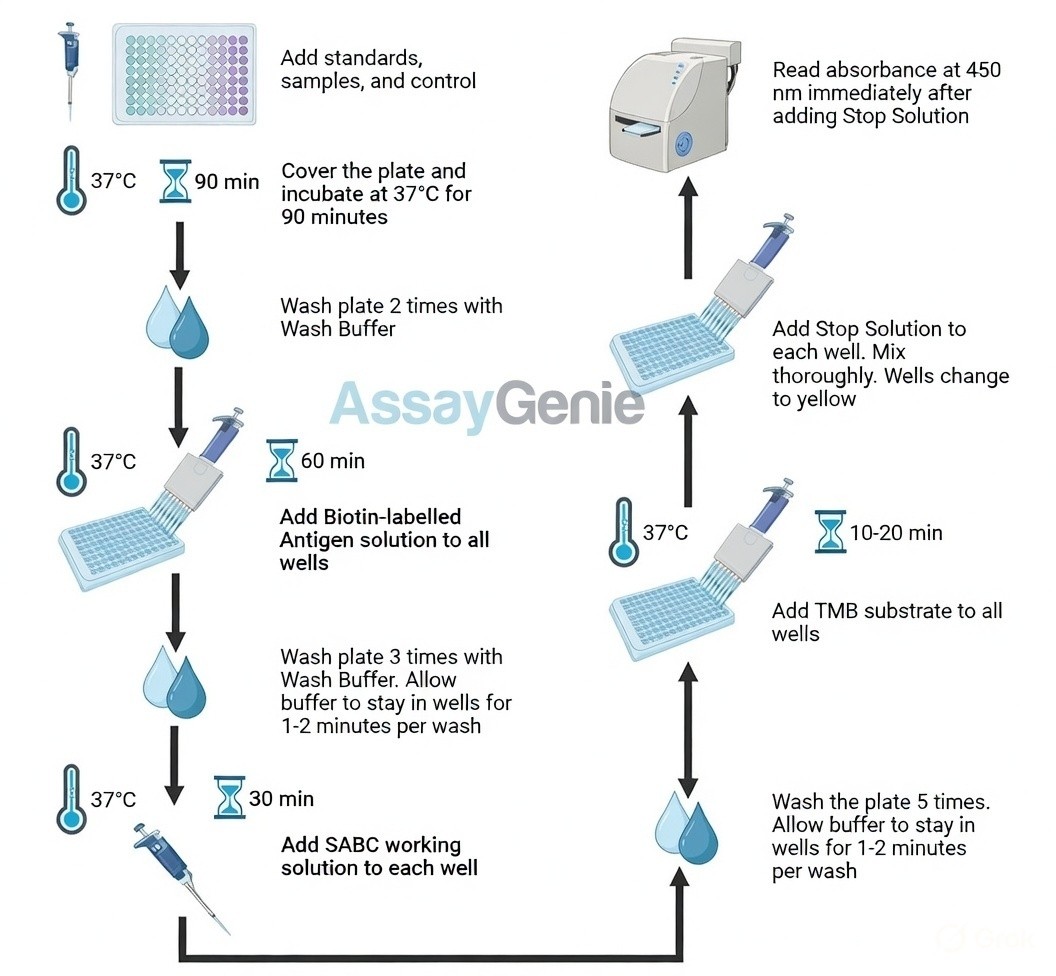
Reagent & Plate Preparation: Equilibrate reagents and TMB substrate to room temperature. Set standard, test sample and control (zero) wells on the pre-coated plate and record their positions.
2
Primary Incubation: Prepare standards, samples, blanks and load into designated wells. Incubate plate at 37°C for 90 minutes to allow antigen binding.
3
Biotin-Labeled Antigen Binding: Add biotin-labeled antigen working solution and incubate at 37°C for 60 minutes.
4
HRP-Streptavidin Binding: Add SABC working solution and incubate at 37°C for 30 minutes.
5
Color Development: Add TMB substrate and incubate in the dark for 10–20 minutes until color develops.
6
Stop Reaction & Reading: Add stop solution and read absorbance at 450 nm immediately.
Sample Type
Protocol
Serum
Allow blood to clot, centrifuge at 1000 × g for 20 minutes and collect supernatant.
Plasma
Collect in anticoagulant tubes, centrifuge at 1000 × g for 15 minutes at 2–8°C and collect plasma.
Tissue Homogenate
Homogenize tissue with PBS or lysis buffer, centrifuge and collect supernatant.
Cell Culture Supernatant
Centrifuge at 2500 rpm for 5 minutes and collect clarified supernatant.
Cell Lysate
Lyse cells on ice using lysis buffer, centrifuge and collect supernatant.
Other Sample Types
For more information about how to process other sample types, (e.g., body fluids, breast milk & more), please contact our Tech Support Team at techsupport@assaygenie.com.
Component
Quantity
Storage
48T
96T
ELISA Microplate (Dismountable)
8×6
8×12
Place the test strips into a sealed foil bag with the desiccant. Store for 1 month at 2-8°C; Store for 12 months at -20°C.
Lyophilized Standard
1 vial
2 vial
Place the standards into a sealed foil bag with the desiccant. Store for 1 month at 2-8°C; Store for 12 months at -20°C.
 ELISA Kit (MOFI01271)")
 ELISA Kit (MOFI01271)")
 ELISA Kit (MOFI01271)")
 ELISA Kit (MOFI01271)")
 ELISA Kit (AEFI00755)")
 ELISA Kit (AEFI00755)")
 ELISA Kit (AEKE12281)")
 ELISA Kit (AEFI00965)")
 ELISA Kit (AEFI00965)")
 ELISA Kit (AEKE11777)")
 ELISA Kit (AEFI01676)")
 ELISA Kit (AEFI01676)")