The RIPK1/RIP Monoclonal Antibody (CAB19580) is a high-quality antibody developed for reliable detection and analysis of target proteins. This antibody, produced using rabbit monoclonal technology, is highly specific and sensitive, making it ideal for use in various applications such as Western blot, immunohistochemistry, and immunofluorescence.RIPK1 is a key player in the regulation of cell death pathways, including apoptosis, necroptosis, and inflammation. Dysregulation of RIPK1 has been implicated in various diseases, including cancer, neurodegenerative disorders, and inflammatory conditions.
This antibody is validated for use in WB, IP, ELISA applications and has demonstrated reactivity against Human samples.
Product Name:
RIPK1/RIP Monoclonal Antibody
SKU:
CAB19580
Size:
20μL, 100μL
Reactivity:
Human
Clone Number:
ARC0059
Conjugate:
Unconjugated
Immunogen:
Recombinant protein (or fragment).This information is considered to be commercially sensitive.
0.5μg-4μg antibody for 200μg-400μg extracts of whole cells
ELISA
Recommended starting concentration is 1 μg/mL. Please optimize the concentration based on your specific assay requirements.
Synonyms:
RIP, RIP1, AIEFL, IMD57, RIP-1, RIPK1/RIP
Positive Sample:
Raji, HeLa, 293T
Cellular Localization:
Cell Membrane, Cytoplasm.
Calculated MW:
76kDa
Observed MW:
76kDa
This gene encodes a member of the receptor-interacting protein (RIP) family of serine/threonine protein kinases. The encoded protein plays a role in inflammation and cell death in response to tissue damage, pathogen recognition, and as part of developmental regulation. RIPK1/RIPK3 kinase-mediated necrosis is referred to as necroptosis. Genetic disruption of this gene in mice results in death shortly after birth.
Purification Method
Affinity purification
Gene ID
8737
RRID
AB_2862680
Buffer Information
Store at -20℃. Avoid freeze / thaw cycles. Buffer: PBS containing 50% glycerol and 0.05% BSA, preserved with proclin300 or sodium azide, pH 7.3.
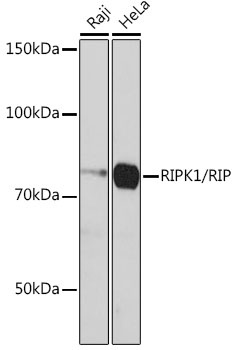
Western blot analysis of various lysates using [KD Validated] RIPK1/RIP Rabbit mAb (CAB19580) at 1:1000 dilution incubated overnight at 4℃. Secondary antibody: HRP-conjugated Goat anti-Rabbit IgG (H+L) (CABS014) at 1:10000 dilution. Lysates/proteins: 25μg per lane. Blocking buffer: 3% nonfat dry milk in TBST. Detection: ECL Basic Kit (AbGn00020). Exposure time: 3min.
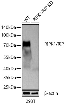
Western blot analysis of lysates from wild type (WT) and RIPK1/RIP knockdown (KD) 293T cells using [KD Validated] RIPK1/RIP Rabbit mAb (CAB19580) at 1:1000 dilution incubated at room temperature for 1.5 hours. Secondary antibody: HRP-conjugated Goat anti-Rabbit IgG (H+L) (CABS014) at 1:10000 dilution. Lysates/proteins: 25 μg per lane. Blocking buffer: 3% nonfat dry milk in TBST. Detection: ECL Basic Kit (AbGn00020). Exposure time: 45s.