
ELISA High Background Troubleshooting: FBS Matrix Interference
Quick answer
A researcher running a Rat Eotaxin/CCL11 (Eotaxin) sandwich ELISA saw sample readings sit above the top standard and wells turn colour the instant TMB was added — even after 1:2 and 1:5 dilutions. This was not a faulty kit. The fetal bovine serum (FBS) in the conditioned culture medium created a high background: the medium-only signal alone reached more than 60% of the top standard, so no practical dilution could bring readings into a reliable range. The fix is to quantify the matrix with a medium-only blank, then either collect serum-free conditioned medium or switch to cell lysates in an HRP-friendly buffer.
On this page
The problem
A researcher wanted to measure secreted CCL11 (Eotaxin) in conditioned medium from primary rat cells. Because the cells are primary and cannot be maintained without serum, the culture medium contained FBS throughout. When the conditioned medium was assayed, every sample read above the top of the standard curve, and the wells developed strong colour the moment TMB substrate was added. Diluting the samples 1:2 with sample diluent left them off-scale; a 1:5 dilution still sat near the top standard. The signal simply would not come down into a measurable window.
Snapshot of the data
The pattern below is what the raw plate looked like. The FBS-containing medium on its own already produced a very high optical density — a large fraction of the assay's full signal — before any secreted target was added.
| Well / condition | OD (450 nm) | Reads within curve? |
| Buffer blank | 0.08 | Reference |
| Top standard (2000 pg/mL) | 2.75 | Upper anchor |
| Medium + FBS only (matrix blank) | 1.78 | ~65% of top standard |
| Sample, 1:2 dilution | > 3.0 (over-range) | No — off scale |
| Sample, 1:5 dilution | 2.05 | No — near ceiling |
Values are illustrative and rounded to show the pattern, not the exact readings from any single run.
When the medium-only OD was subtracted from the sample ODs, a few replicates dropped back toward the working part of the curve — but with the background this high, the usable window is narrow.
| Replicate | Raw OD | Minus matrix blank | Interpretation |
| A | 2.30 | 0.52 | Falls within curve |
| B | 1.95 | 0.17 | Low, near lower limit |
| C | 2.60 | 0.82 | Within curve, wide spread vs. A/B |
Values are illustrative. Biological and technical replicates varied widely, which is itself a warning sign when the background is this large.
Why it happens
FBS is a rich, undefined protein matrix. In a sandwich ELISA, those serum proteins can bind non-specifically to the plate, the capture antibody, or the detection antibody, and they raise the assay's baseline signal across every well. This is matrix interference, and it is independent of how much target the sample actually contains.
The reason dilution did not rescue the assay is arithmetic. Dilution lowers the target signal and the matrix background together. When the background is a small fraction of the top standard, diluting a saturated sample eventually lands it inside the curve. But when the matrix alone already reaches ~60–65% of the maximum signal, you run out of curve before you run out of background — the sample and its matrix descend in lock-step and never separate into a clean, measurable window.
Our analysis
We asked the researcher to include a medium-only well — the same FBS-containing medium with no cells — as a matrix blank, and to share the plate layout. Subtracting that matrix blank from the sample ODs pulled several replicates back into the reliable region of the standard curve, which confirmed there was real target signal underneath the noise. However, because the background stood at more than 60% of the top standard, the corrected values sat on a compressed, low-confidence part of the curve, and replicates that should have matched diverged. Chasing the answer with further dilution was not going to give trustworthy numbers.
Root cause
Serum-matrix interference, not a defective kit. The high FBS background in the conditioned medium saturated the assay and left almost no dynamic range for the secreted target. Listing "cell culture supernatant" as a compatible sample type does not guarantee that a serum-rich conditioned medium will behave — the specific matrix still has to be checked.
What we recommended
Key takeaway
Off-scale readings and instant TMB colour usually point to background and matrix, not a bad kit. Quantify the background with a matrix-matched blank before you blame the assay — and if a serum-rich sample won't come into range by dilution, change the sample, not just the dilution. For more standard-curve and matrix guidance, see the ELISA Support Hub →
Frequently asked questions
Why didn't diluting the sample fix the off-scale signal?
Because the FBS background was already ~60–65% of the top standard. Dilution lowers the target and the background together, so the sample never separates from its matrix into a clean measurable window — you exhaust the curve before you clear the noise.
Can I just remove FBS for the assay?
Where the cells tolerate it, collect conditioned medium under serum-free or reduced-serum conditions for a defined window before harvest. If the cells genuinely require serum to survive, a cell lysate is usually the better route for a secreted target.
Which lysis buffers are ELISA-safe?
A Tris/NaCl/SDS buffer at pH 7.3 works well. Avoid NP-40, Triton X-100, DTT and standard RIPA formulations, as these inhibit HRP and suppress the colour reaction.
Does "cell culture supernatant" on the datasheet mean my medium will work?
Listed sample types are general compatibility guidance. A serum-rich conditioned medium is a demanding matrix, so always validate it with a matrix blank and, ideally, a linearity-of-dilution check before relying on the numbers.
Related reading
ELISA Controls Guide — matrix & spiked-matrix controls →
Troubleshooting High ELISA Concentrations →
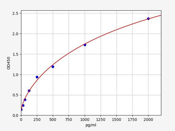
SKU: RTFI00115 · Sandwich ELISA (double antibody)
| Detection range | 31.25–2000 pg/mL |
| Sensitivity | 18.75 pg/mL |
| Sample types | Serum, plasma, cell culture supernatant, cell or tissue lysate |
€699
View product →Fighting high background or off-scale ELISA readings?
Our PhD-level scientific support team can review your data and help you find the right sample prep for your matrix.
This article is based on an anonymised technical-support case and is provided for general educational purposes. Assay values shown are illustrative. Optimal sample preparation and dilution vary by cell type, matrix and target; validate conditions for your own samples before generating final data.
Recent Posts
-
Suspected Plague Death at a Siberian Anti-Plague Institute: What Researchers Should Know
Infectious Disease · News AnalysisSuspected Plague Death at a Siberian Anti-Plague Institute: What …6th Oct 2026 -
LPS ELISA Troubleshooting: The Importance of Sample Nature
Quick answer A researcher measuring circulating lipopolysaccharide (LPS) in human serum …25th Sep 2026 -

Sekundärantikörper wählen: Die Regel, die im Labor wirklich zählt
Sekundärantikörper wählen: Die Regel, die im Labor wirklich zählt Wählen Sie einen Sekundärantikörp …24th Sep 2026