
What Are Oligodendrocytes? Functions, Markers & Disease Links
Oligodendrocytes: Myelination, Lineage Markers and Disease
Oligodendrocytes are the myelinating glia of the central nervous system. A single oligodendrocyte can wrap segments of dozens of different axons, and the myelin it produces is what makes rapid saltatory conduction possible. They also feed axons metabolically. This guide covers the lineage from progenitor to mature cell, the markers used to identify each stage, how they differ from Schwann cells, and the diseases in which they fail.
Browse myelin & glial markers →Key takeaways
- Oligodendrocytes myelinate axons in the CNS. One cell myelinates segments of many different axons — typically forty to fifty internodes — whereas one Schwann cell myelinates a single internode on a single axon.
- Both cell types produce multilamellar myelin of many concentric wraps. The difference is how many axons each serves, not how many layers it lays down.
- Myelin enables saltatory conduction, with action potentials regenerating only at the nodes of Ranvier between sheaths.
- Oligodendrocytes also provide metabolic support, delivering lactate and pyruvate to axons through monocarboxylate transporters — a role independent of insulation, and one whose failure causes axon degeneration.
- The lineage runs OPC → pre-oligodendrocyte → immature → mature, with distinct markers at each stage. NG2 and PDGFR-alpha mark progenitors; MBP, PLP and CNPase mark mature myelinating cells.
- OPCs persist throughout adult life and are the largest proliferating cell population in the adult CNS, which is what makes remyelination possible in principle.
- Multiple system atrophy is the neurodegenerative disease centred on oligodendrocytes, defined by alpha-synuclein inclusions within them.
Contents
- What oligodendrocytes are
- Functions: insulation and metabolic support
- The oligodendrocyte lineage
- Lineage markers by stage
- Oligodendrocytes versus Schwann cells
- Histology and imaging techniques
- Diseases involving oligodendrocytes
- Remyelination and why it fails
- Treatment strategies
- Choosing markers
- Frequently asked questions
Oligodendrocyte and glial markers
Transcription factors for the lineage, the three principal myelin proteins, and GFAP to distinguish astrocytes in mixed glial preparations.
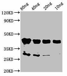
OLIG2 Antibody
The defining lineage transcription factor — expressed from progenitor through to mature oligodendrocyte.
View product →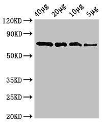
SOX10 Antibody
Maintained across the whole lineage and also expressed by Schwann cells, so pair with a CNS context.
View product →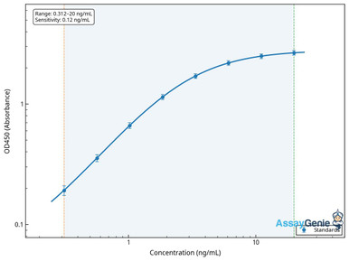
Human Myelin Basic Protein (MBP) ELISA Kit
The most abundant myelin protein; released into CSF and serum during active demyelination.
View product →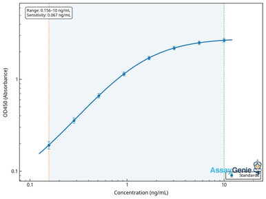
Human Myelin Proteolipid Protein (PLP1) ELISA Kit
The major structural myelin protein; PLP1 mutations cause Pelizaeus-Merzbacher disease.
View product →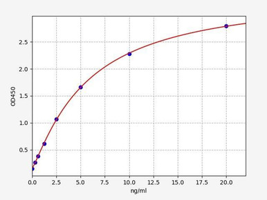
Human CNPase ELISA Kit
An early myelination marker, expressed before MBP and useful for staging differentiation.
View product →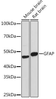
GFAP Rabbit Polyclonal Antibody
The astrocyte marker — essential as a counterstain when identifying oligodendrocytes in mixed cultures.
View product →What oligodendrocytes are

Oligodendrocytes are one of the four major glial cell types of the central nervous system, alongside astrocytes, microglia and ependymal cells. Their defining role is to produce myelin — the lipid-rich multilayered membrane that insulates axons.
The name means “cell with few branches”, which undersells them: each cell extends processes to reach many separate axons, and the myelin membrane a single oligodendrocyte produces can exceed a hundred times the surface area of its own cell body.
They are found in both white matter, where myelinated tracts predominate, and grey matter, where satellite oligodendrocytes sit adjacent to neuronal cell bodies.
Functions: insulation and metabolic support

Saltatory conduction
Myelin raises membrane resistance and lowers capacitance, so current spreads passively along the internode rather than depolarising every point of membrane. Action potentials regenerate only at the nodes of Ranvier — short unmyelinated gaps densely packed with voltage-gated sodium channels.
The result is conduction that jumps node to node, an order of magnitude faster than an unmyelinated axon of the same diameter and far more energy-efficient, because sodium entry is restricted to a small fraction of the membrane.
Metabolic support of axons
This is the function most often omitted, and it is not secondary. Oligodendrocytes deliver lactate and pyruvate to the axons they ensheath, through monocarboxylate transporters, supplying substrate for axonal mitochondria.
The evidence that this matters independently of insulation is direct: disrupting oligodendrocyte metabolic support causes axon degeneration even where the myelin sheath remains structurally intact. It reframes the oligodendrocyte as a metabolic partner of the axon rather than simply its insulator — and it is one reason axons are lost in chronic demyelinating disease.
The oligodendrocyte lineage
Oligodendrocytes develop through a defined sequence, and each stage is morphologically and molecularly distinct.
| Stage | Behaviour | Morphology |
|---|---|---|
| Oligodendrocyte progenitor cell (OPC) | Migratory and proliferative | Bipolar, few processes |
| Pre-oligodendrocyte | Migration ceases; proliferation slows | Multipolar, more processes |
| Immature oligodendrocyte | Contacts axons but does not yet myelinate | Highly branched |
| Mature myelinating oligodendrocyte | Post-mitotic; produces and maintains myelin | Extensive membrane sheets wrapping axons |
During development, OPCs arise in successive waves from distinct ventricular zone domains and migrate throughout the CNS. Crucially, OPCs persist into adulthood, remaining as a dispersed population that continues to divide — they are the largest proliferating cell population in the adult brain, and they are the reservoir from which remyelination proceeds.
Not every axon contact leads to myelination. Oligodendrocytes select targets by diameter and by neuronal activity, so myelination is responsive to use rather than fixed.
Lineage markers by stage
Markers should be chosen for the stage being studied, since several are expressed across the whole lineage and cannot distinguish progenitors from mature cells.
| Marker | Stage | Notes |
|---|---|---|
| OLIG1 / OLIG2 | Whole lineage | Transcription factors required for specification; OLIG2 is the standard lineage label |
| SOX10 | Whole lineage | Also expressed by Schwann cells, so interpret with CNS context |
| NG2 (CSPG4) | OPC | Together with PDGFR-alpha, the standard progenitor marker pair |
| PDGFR-alpha | OPC | Lost as cells exit the progenitor state |
| O4 | Pre-oligodendrocyte onward | Marks the transition out of the progenitor stage |
| CNPase | Immature and mature | An early myelination marker, appearing before MBP |
| MBP | Mature, myelinating | The most abundant myelin protein |
| PLP1 | Mature, myelinating | The major structural myelin protein |
| MOG | Mature, late | Surface-exposed and the target antigen in MOG antibody-associated disease |
A note on nomenclature: there is no marker called “Olig-17”, a term that appears in some secondary sources. The OLIG family comprises OLIG1, OLIG2 and OLIG3, and the genuine progenitor markers are NG2 and PDGFR-alpha.
Oligodendrocytes versus Schwann cells
| Feature | Oligodendrocyte | Schwann cell |
|---|---|---|
| Location | Central nervous system | Peripheral nervous system |
| Axons served | Many — typically 40 to 50 internodes across different axons | One internode on one axon |
| Myelin structure | Multilamellar, many concentric wraps | Multilamellar, many concentric wraps |
| Developmental origin | Neuroepithelium of the ventricular zones | Neural crest |
| Basal lamina | Absent | Present, surrounding the cell |
| Regeneration support | Poor — CNS myelin debris inhibits axon regrowth | Good — Schwann cells guide regenerating axons |
Two points are commonly muddled. First, both produce multilamellar myelin; Schwann cell myelin is not a single layer. The real distinction is one-to-many versus one-to-one. Second, the regeneration difference is clinically significant — peripheral nerves can recover after injury in a way central tracts largely cannot, and Schwann cells are a major reason why.
Histology and imaging techniques
- Luxol fast blue stains myelin directly and remains the standard histochemical method for assessing demyelination in tissue sections.
- Haematoxylin and eosin shows general morphology; oligodendrocytes appear as small cells with round dark nuclei and a characteristic perinuclear halo in fixed tissue.
- Immunohistochemistry and immunofluorescence allow stage-specific identification using the markers above, and permit co-labelling with GFAP or IBA1 to distinguish astrocytes and microglia.
- Electron microscopy resolves individual myelin lamellae and is used to measure g-ratio, the ratio of axon diameter to fibre diameter, which quantifies myelin thickness.
- MRI assesses myelin in vivo, with magnetisation transfer and diffusion measures used to track white matter change over time.
Diseases involving oligodendrocytes
| Category | Examples | Mechanism | |
|---|---|---|---|
| Inflammatory demyelination | Multiple sclerosis; MOG antibody-associated disease | Immune attack on myelin and oligodendrocytes, with axon loss following | undefined |
| Oligodendrocyte synucleinopathy | Multiple system atrophy | Alpha-synuclein accumulates as glial cytoplasmic inclusions within oligodendrocytes — the defining lesion | undefined |
| Inherited leukodystrophy | Pelizaeus-Merzbacher (PLP1); metachromatic leukodystrophy; adrenoleukodystrophy | Defective myelin protein or lipid metabolism | undefined |
| Hypoxic-ischaemic injury | Periventricular leukomalacia in preterm infants | Immature OPCs are selectively vulnerable to oxidative and excitotoxic injury | undefined |
| Tumours | Oligodendroglioma | Glioma of oligodendroglial lineage, defined by IDH mutation with 1p/19q codeletion | undefined |
Two clarifications are worth making. Multiple system atrophy, not Alzheimer’s or Parkinson’s disease, is the neurodegenerative condition centred on oligodendrocytes; while oligodendrocyte changes are an active research interest in the commoner dementias, the causal picture there is not established.
And oligodendrogliomas are not usefully described as aggressive. Those defined by IDH mutation with 1p/19q codeletion are the most chemosensitive and favourable-prognosis diffuse gliomas, with survival often measured in many years — the molecular classification matters more than the histology.
Remyelination and why it fails
Remyelination is possible because adult OPCs persist. It occurs spontaneously in early multiple sclerosis, producing shadow plaques — areas of thin but genuine new myelin.
It becomes progressively less effective with age and chronicity, and the reasons are reasonably well defined:
- Differentiation block. OPCs are recruited to chronic lesions but arrest before maturing, so the limiting step is differentiation rather than progenitor supply.
- Inhibitory debris. Myelin fragments contain molecules that inhibit OPC differentiation, and clearance by microglia becomes less efficient with age.
- Astrocytic scarring creates a physical and chemical barrier to OPC migration.
- Ageing reduces the intrinsic differentiation capacity of OPCs themselves.
That the block is at differentiation rather than recruitment is why remyelination research concentrates on agents that push already-present OPCs to mature.
Treatment strategies
Research context only — no dosing is given and this is not clinical guidance.
- Immunomodulation. In multiple sclerosis, disease-modifying therapies reduce relapse frequency and new lesion formation. They act on the immune attack rather than restoring lost myelin.
- Remyelination strategies. Aimed at releasing the OPC differentiation block; several repurposed compounds have reached clinical trials, with mixed results so far.
- Cell transplantation. OPC or stem cell grafting, investigated particularly for leukodystrophies where the host cells are genetically defective.
- Neuroprotection. Preserving axons independently of remyelination, relevant because axon loss rather than demyelination drives lasting disability.
- Symptomatic and rehabilitative care, which remains central to function and quality of life.
Choosing markers
OLIG2 and SOX10 for the lineage, MBP, PLP1 and CNPase for myelination stage, and GFAP to separate astrocytes in mixed glial preparations.
Browse myelin & glial markers →Frequently asked questions
What is the difference between oligodendrocytes and Schwann cells?
Oligodendrocytes myelinate in the CNS and each one serves segments of many axons — typically forty to fifty internodes. A Schwann cell myelinates a single internode on a single axon in the PNS. Both produce multilamellar myelin; the difference is coverage, not layer count.
Do Schwann cells make only one layer of myelin?
No. Schwann cell myelin consists of many concentric wraps, just like CNS myelin. The frequent claim that it is a single layer confuses the number of axons served with the number of membrane layers.
Which markers identify oligodendrocyte progenitors?
NG2 (CSPG4) and PDGFR-alpha. OLIG2 and SOX10 are expressed across the entire lineage and cannot distinguish progenitors from mature cells, so they should be paired with a stage-specific marker.
Is there a marker called Olig-17?
No. The OLIG family consists of OLIG1, OLIG2 and OLIG3. “Olig-17” appears in some secondary sources but does not correspond to a real protein — the progenitor markers are NG2 and PDGFR-alpha.
Do oligodendrocytes do anything besides insulate axons?
Yes. They supply axons with lactate and pyruvate through monocarboxylate transporters. Disrupting this metabolic support causes axon degeneration even when the myelin sheath is structurally intact.
Which neurodegenerative disease most directly involves oligodendrocytes?
Multiple system atrophy, defined by alpha-synuclein inclusions inside oligodendrocytes. Oligodendrocyte involvement in Alzheimer’s and Parkinson’s disease is an area of research but is not established as causal.
Why does remyelination fail in chronic multiple sclerosis?
Chiefly because recruited progenitors arrest before maturing. Inhibitory myelin debris, astrocytic scarring and reduced intrinsic capacity with age all contribute, which is why therapeutic effort focuses on driving differentiation rather than supplying more progenitors.

Marina Alberto, PhD, holds a robust academic background in Biotechnology, earning her Bachelor’s Degree and PhD in Science and Technology from Quilmes National University. Her research spans cancer immunotherapy, glycan profiling, and vaccine development, including innovative projects on pediatric leukemia diagnosis and cancer-associated carbohydrate-mimetic vaccines. She currently serves as a Technical Support and Sales Specialist at Assay Genie.
Recent Posts
-
Suspected Plague Death at a Siberian Anti-Plague Institute: What Researchers Should Know
Infectious Disease · News AnalysisSuspected Plague Death at a Siberian Anti-Plague Institute: What …6th Oct 2026 -
LPS ELISA Troubleshooting: The Importance of Sample Nature
Quick answer A researcher measuring circulating lipopolysaccharide (LPS) in human serum …25th Sep 2026 -

Sekundärantikörper wählen: Die Regel, die im Labor wirklich zählt
Sekundärantikörper wählen: Die Regel, die im Labor wirklich zählt Wählen Sie einen Sekundärantikörp …24th Sep 2026