
Cell Synchronisation Methods
Cell Synchronisation Methods: Protocols for G1, S, G2 and M Phase Arrest
An asynchronous culture contains cells at every stage of the cycle at once, so any measurement is an average across phases. Synchronisation forces cells into one phase so that phase-specific events can actually be observed. This guide covers the chemical and physical approaches, six working protocols with concentrations and timings, how to confirm synchrony by flow cytometry, and the artefacts these methods introduce.
Browse cell cycle reagents →Key takeaways
- Synchronisation is achieved either by chemical blockade — arresting cells at a defined point — or by physical fractionation, which selects cells already in a phase without perturbing them.
- Double thymidine block arrests at the G1/S boundary; nocodazole and taxol arrest in mitosis; RO-3306 arrests at G2/M by inhibiting CDK1.
- Chemical methods are efficient but not innocuous — they impose stress, and cells released from a block are not equivalent to cells that reached that phase naturally.
- Thymidine blocks work by nucleotide pool imbalance, so a single block leaves cells spread through S phase; the second block is what produces a tight G1/S population.
- Nocodazole and taxol both arrest in mitosis but by opposite means — nocodazole depolymerises microtubules, taxol hyper-stabilises them.
- Always confirm synchrony rather than assuming it: fix, treat with RNase, stain DNA with propidium iodide and read the DNA content profile by flow cytometry.
- Prolonged mitotic arrest triggers apoptosis or mitotic slippage, so arrest duration is a real experimental variable and not a free parameter.
Contents
- What is cell synchronisation?
- Stages of the cell cycle
- Chemical blockade and physical fractionation
- Which agent for which phase
- Protocol: double thymidine block
- Protocol: nocodazole mitotic arrest
- Protocol: CDK inhibitors after synchronisation
- Protocol: taxol-arrested cells with CDK inhibitors
- Protocol: RO-3306 G2/M arrest
- Confirming synchronisation
- Pitfalls and artefacts
- Choosing reagents
- Frequently asked questions
Reagents for cell synchronisation
The panel below covers the lysis buffer used throughout these protocols, the DNA stain for confirming synchrony, and the markers that report which phase cells are actually in.

GenieLyse RIPA Lysis Buffer
Used in four of the protocols below to prepare whole cell extracts after release from arrest.
View product →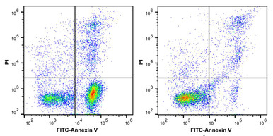
Propidium Iodide Staining Solution
Stains DNA stoichiometrically — the basis of the DNA content profile that confirms synchrony.
View product →
Anti-Phospho-Histone H3 (Ser10) Monoclonal Antibody
Serine 10 phosphorylation is the definitive mitotic marker, distinguishing true M phase from G2.
View product →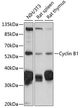
Cyclin B1 Rabbit Polyclonal Antibody
Accumulates through G2, peaks at mitosis and is destroyed at anaphase — a direct readout of position in the cycle.
View product →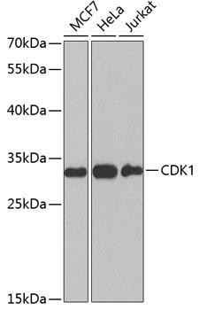
CDK1 Rabbit Polyclonal Antibody
The kinase RO-3306 inhibits; CDK1-cyclin B activity is what drives entry into mitosis.
View product →
EdU Cell Proliferation Flow Cytometry Assay Kit
Labels newly synthesised DNA, identifying S-phase cells directly rather than inferring from DNA content.
View product →What is cell synchronisation?
In a normal culture, cells occupy every stage of the cycle simultaneously. Any measurement taken from that population is therefore an average across phases, which obscures anything phase-specific. Cell synchronisation brings the population to a common point so that events tied to a particular phase can be observed directly, and so that progression can be followed as a cohort after release.
It underpins work on cell cycle regulation, on the timing of protein expression and degradation, on DNA replication and repair, and on how cytotoxic and targeted drugs act at particular phases.
Stages of the cell cycle
The cycle comprises four active phases plus a quiescent state, and synchronisation targets the transitions between them.
| Phase | What happens |
|---|---|
| G0 | Quiescence — cells are viable and metabolically active but not committed to dividing. Entered on serum withdrawal or contact inhibition. |
| G1 | Growth and preparation for DNA synthesis. The restriction point late in G1 is the commitment step, after which the cell proceeds independently of mitogens. |
| S | DNA replication. Cells here have between 2n and 4n DNA content, which is what makes them identifiable by flow cytometry. |
| G2 | Growth and checking of replicated DNA before division. CDK1-cyclin B accumulates but is held inactive. |
| M | Mitosis — chromosome segregation and cytokinesis. The shortest phase, typically under an hour. |
For synchronisation purposes the useful boundaries are G1/S, where thymidine acts, G2/M, where CDK1 inhibition acts, and mitosis itself, where microtubule agents act.
Chemical blockade and physical fractionation
Chemical blockade
Chemical methods arrest cells at a defined point by blocking a required process. Thymidine, aminopterin, hydroxyurea and cytosine arabinoside all interfere with DNA synthesis and arrest cells in or at the entry to S phase. Serum starvation for around 24 hours drives cells into G0/G1, and the effect is reversed by restoring serum. CDK inhibitors and microtubule agents arrest later in the cycle.
The advantage is efficiency — a high proportion of cells end up in one phase. The cost is that arrest is a stress: cells are held at a checkpoint rather than passing through it naturally, and the longer they are held, the more they diverge from unperturbed cells.
Physical fractionation
Physical methods separate cells that are already in a given phase, using differences in size, density, surface epitopes or light scatter. Centrifugal elutriation separates on size and sedimentation velocity; fluorescence-activated cell sorting separates on scatter or on fluorescence from stained DNA, RNA, protein or surface antigens.
The advantage is that no drug is applied, so cells are physiologically unperturbed. The costs are lower yield, lower purity, and the requirement for specialised equipment. Where the biology being studied is itself sensitive to checkpoint stress, fractionation is the more defensible choice despite those costs.
Which agent for which phase
| Target phase | Agents used |
|---|---|
| G1 arrest | Double thymidine block; serum starvation; CDK inhibition |
| G2 arrest | Microtubule inhibition; CDK inhibition |
| G2/M arrest | RO-3306 (selective CDK1 inhibitor) |
| M phase | Taxol (paclitaxel); nocodazole |
The mechanisms differ in ways that matter. Thymidine works by imbalancing the deoxynucleotide pool, which stalls replication forks. Nocodazole depolymerises microtubules, preventing spindle assembly; taxol does the opposite, stabilising microtubules so they cannot undergo the dynamic instability mitosis requires. Both trigger the spindle assembly checkpoint and arrest cells in mitosis, but the spindle states are entirely different — which matters if the readout involves microtubules.
Protocol: double thymidine block
Arrests cells at the G1/S boundary. The double block exists because a single thymidine exposure leaves cells distributed through S phase; the release period lets them run to completion, and the second block then catches the whole population at the boundary.
| Step | Procedure |
|---|---|
| 1. | To synchronise cells at the G1/S border a freshly prepared thymidine solution (16 mM) was made up in complete medium and filter-sterilised using a 0.22 µm filter disc. |
| 2. | Cells (1 × 10⁷) were cultured in 60 ml complete medium containing thymidine (2 mM) for 16 h in a 175 cm2 cell culture flask. |
| 3. | The cells were then resuspended in complete medium (52.5 ml) for 8 h to allow cells to reenter the cell cycle. |
| 4. | Cells were harvested by centrifugation at 400 × g for 5 min and washed twice in complete medium (10 ml). Cells (1 × 10⁷) were cultured in 60 ml complete medium containing thymidine (2mM) for 16 h to synchronise cells at the G1/S border. |
| 5. | Cell synchronisation was confirmed by flow cytometry. |
Protocol: nocodazole mitotic arrest
Arrests cells in mitosis by preventing spindle formation. The timed release samples below allow mitotic exit to be followed as a cohort.
| Step | Procedure |
|---|---|
| 1. | Cells (1 × 10⁷) were synchronised in mitosis by treating cells for 18 h with Nocodazole (100 nM) and incubated at 37 °C in a 175 cm2 |
| 2. | Cells were washed with complete medium three times and re-plated in complete medium and samples were taken at 0, 30, 60, 120 and 180 min post-release. |
| 3. | As a control, one sample was re-plated in the presence of Nocodazole (100 nM) to maintain cells in mitosis and a sample was taken 180 min post-release. |
| 4. | Cells were lysed in RIPA buffer and whole cell extracts were prepared. Samples were stored at -20 °C. |
Protocol: CDK inhibitors after synchronisation
Applied to K562 cells first arrested by double thymidine block, this protocol compares several kinase inhibitors and a proteasome inhibitor in taxol-arrested mitotic cells.
| Step | Procedure |
|---|---|
| 1. | K562 cells were arrested in G1/S phase by double thymidine block (DTB) as previously described (see first sample protocol) |
| 2. | Cells were harvested by centrifugation at 400 × g for 5 min and washed twice in complete medium (10 ml). |
| 3. | The cells (1 × 10⁶/ ml) were then resuspended in complete medium and allowed to rest for 1 h. |
| 4. | Cells were treated with either vehicle (0.1% (v/v) DMSO) or Taxol (1 µM) for 11 h. 12 h post DTB cells were treated with either vehicle (0.1% (v/v) DMSO) or with the Kinase inhibitors, BI2536 (10 µM) Roscovitine (20 µM), RO-3306 (9 µM), Purvalanol A (10 µM) the proteasome inhibitor, MG-132 (10 µM) for 2 h. |
| 5. | Cells were lysed with RIPA buffer and whole cell extracts were prepared. |
| 6. | Samples were stored at -20 °C. |
Note the inhibitor panel: BI2536 targets Polo-like kinase 1, roscovitine and purvalanol A are broad CDK inhibitors, RO-3306 is CDK1-selective, and MG-132 blocks the proteasome — which prevents cyclin B destruction and so prevents mitotic exit.
Protocol: M-phase treatment of arrested cells
A shorter variant comparing taxol and nocodazole arrest, followed by CDK inhibition with or without proteasome inhibition.
| Step | Procedure |
|---|---|
| 1. | Cells (1 × 10⁶) were treated with either Vehicle (0.1% (v/v) DMSO), Taxol (1 µM) or Nocodazole (1 µM) for 12 h. |
| 2. | The cells were treated with the kinase inhibitor Roscovitine (20 µM) or Roscovitine plus the proteasome inhibitor, MG-132 (10 µM) for 2 h. Cells were harvested by centrifugation at 400 × g for 5 min and lysed with 30 µl RIPA buffer. |
| 3. | Samples were stored at -20 °C. |
Protocol: RO-3306 G2/M arrest
RO-3306 is a selective CDK1 inhibitor, so it holds cells at the G2/M boundary rather than within mitosis. Because the block is immediately upstream of mitotic entry, washout produces an unusually synchronous wave of cells entering mitosis — which is what the timed sampling below captures.
| Step | Procedure |
|---|---|
| 1. | K562 cells were treated with either vehicle (0.1% (v/v) DMSO) or RO-3306 (9 µM) for 18 h. |
| 2. | Cells were washed three times in complete medium and re-plated in 10 ml of complete medium treated with either vehicle (0.1% (v/v) DMSO) or MG-132 (10 µM). |
| 3. | Samples were taken at 0, 15, 30, 45, 60 and 90 min post-wash. |
| 4. | K562 cells were lysed with RIPA buffer and whole cell extracts were prepared. |
| 5. | Samples were stored at -20 °C. |
Confirming synchronisation
Synchrony should be measured, not assumed. The standard approach reads DNA content by flow cytometry: G0/G1 cells have 2n DNA, G2/M cells have 4n, and S phase cells fall between.
| Step | Procedure |
|---|---|
| 1. | Fix and permeabilize your cells in 70 % ethanol |
| 2. | Stain with 40 µg/ml propidium iodide, and include 25 µg/ml of RNase (to degrade RNA and ensure that you stain DNA only). |
| 3. | Run your samples on the flow cytometer. |
Two points on the method. The RNase step is not optional — propidium iodide binds double-stranded RNA as well as DNA, so without it the profile is distorted. And DNA content alone cannot distinguish G2 from M, since both are 4n; separating them requires a mitotic marker such as phospho-histone H3 at serine 10.
Complementary readouts include cyclin B1, which accumulates through G2 and is destroyed at anaphase, and EdU incorporation, which labels cells actively synthesising DNA rather than inferring S phase from DNA content.
Pitfalls and artefacts
| Issue | Why it happens | What to do |
|---|---|---|
| Synchrony decays after release | Cells progress at slightly different rates, so the cohort spreads with each hour | Sample early; do not expect synchrony to survive a full cycle |
| Arrest itself alters the biology | Checkpoint activation changes transcription, signalling and protein stability | Include a released-but-untreated control; consider fractionation if the readout is checkpoint-sensitive |
| Thymidine toxicity | Prolonged nucleotide imbalance causes replication stress and DNA damage | Keep to established block and release timings rather than extending them |
| Mitotic slippage | Cells held in mitosis degrade cyclin B slowly and exit without dividing, becoming 4n G1 cells | Limit arrest duration; verify mitotic status with phospho-histone H3 rather than DNA content |
| Apoptosis during prolonged arrest | Extended mitotic arrest activates cell death pathways | Use the shortest effective arrest; monitor viability alongside synchrony |
| Cell-line variability | Doubling times and drug sensitivities differ substantially between lines | Optimise concentration and duration for each line rather than transferring conditions |
The general caution is worth stating plainly: a chemically synchronised population is not simply a normal population caught at one moment. It is a population that has been held at a checkpoint, and for some questions that difference is the experiment.
Choosing reagents
RIPA lysis buffer and propidium iodide for these protocols, plus phospho-histone H3, cyclin B1 and CDK1 antibodies and EdU labelling for confirming which phase your cells are actually in.
Browse cell cycle reagents →Frequently asked questions
Why is a double thymidine block needed rather than a single one?
Because a single exposure arrests cells wherever they are within S phase, leaving them spread across it. Releasing lets that cohort finish S phase, and the second block then catches essentially the whole population at the G1/S boundary.
What is the difference between nocodazole and taxol arrest?
Both arrest cells in mitosis by triggering the spindle assembly checkpoint, but by opposite mechanisms — nocodazole depolymerises microtubules while taxol hyper-stabilises them. The spindle states differ completely, which matters if your readout involves microtubules.
How do I confirm cells are synchronised?
Fix and permeabilise in 70% ethanol, treat with RNase, stain DNA with propidium iodide and read DNA content by flow cytometry. G0/G1 cells are 2n, G2/M cells 4n, S phase in between.
Why is RNase needed for propidium iodide staining?
Because propidium iodide binds double-stranded RNA as well as DNA. Without RNase digestion the signal includes an RNA contribution and the DNA content profile is distorted.
Can flow cytometry distinguish G2 from mitosis?
Not on DNA content alone — both are 4n. Separating them requires a mitosis-specific marker, most commonly phosphorylation of histone H3 at serine 10.
What is mitotic slippage?
Cells held in mitotic arrest slowly degrade cyclin B and exit mitosis without dividing, ending up as tetraploid G1 cells. It is a common artefact of prolonged arrest and one reason to keep arrest as short as the experiment allows.
Is chemical synchronisation equivalent to natural cell cycle progression?
No. Arrest holds cells at a checkpoint and imposes stress that alters transcription, signalling and protein stability. Where that matters, physical fractionation is unperturbing, at the cost of lower yield and purity.
Recent Posts
-
Suspected Plague Death at a Siberian Anti-Plague Institute: What Researchers Should Know
Infectious Disease · News AnalysisSuspected Plague Death at a Siberian Anti-Plague Institute: What …6th Oct 2026 -
LPS ELISA Troubleshooting: The Importance of Sample Nature
Quick answer A researcher measuring circulating lipopolysaccharide (LPS) in human serum …25th Sep 2026 -

Sekundärantikörper wählen: Die Regel, die im Labor wirklich zählt
Sekundärantikörper wählen: Die Regel, die im Labor wirklich zählt Wählen Sie einen Sekundärantikörp …24th Sep 2026