
Phosphorylation: Types, ATP Synthesis & Cell Signalling [Guide]
Phosphorylation: Types, ATP Synthesis and Cell Signalling
Adding a phosphate group is how cells both store energy and switch proteins on and off. The same chemistry underlies ATP synthesis in mitochondria and chloroplasts and the regulatory phosphorylation that drives every signalling cascade. This guide covers the three metabolic routes to ATP, how protein phosphorylation works as a switch, and how phosphorylation is measured.
Browse phospho antibodies →Key takeaways
- Phosphorylation transfers a phosphoryl group to a molecule, and because phosphatases reverse it, the modification works as a switch rather than a permanent change.
- Three metabolic routes generate ATP: substrate-level phosphorylation in the cytosol, oxidative phosphorylation in mitochondria, and photophosphorylation in chloroplasts.
- Substrate-level phosphorylation transfers phosphate directly from a high-energy substrate to ADP and needs neither oxygen nor an electron transport chain.
- Oxidative phosphorylation is indirect: electron transport builds a proton gradient, and ATP synthase converts that gradient into ATP.
- The familiar figure of 36–38 ATP per glucose is outdated — current estimates are around 30 to 32, because the proton-to-ATP ratio is non-integral and transport carries a cost.
- In proteins, phosphorylation occurs mainly on serine, threonine and tyrosine, with serine by far the most common.
- Because phosphorylation changes state rather than abundance, it must be measured with phospho-specific reagents and normalised to total protein.
Contents
- The phosphoryl group
- The three types of phosphorylation
- Substrate-level phosphorylation
- Oxidative phosphorylation
- Oxidative versus substrate-level
- How much ATP does glucose actually yield?
- Photophosphorylation
- Protein phosphorylation as a switch
- Measuring phosphorylation
- Choosing phospho reagents
- Frequently asked questions
Reagents for phosphorylation studies
Phosphorylation is a state, not an amount, so the reagents that matter are phospho-specific. The panel covers the three phosphorylatable residues, ATP as the product, and a worked phospho-plus-total pair.
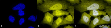
Anti-Phosphoserine Antibody
Detects phosphoserine across the proteome — the most abundant phospho-residue by a wide margin.
View product →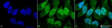
Anti-Phosphothreonine Antibody
Detects phosphothreonine, the second most common site of serine/threonine kinase activity.
View product →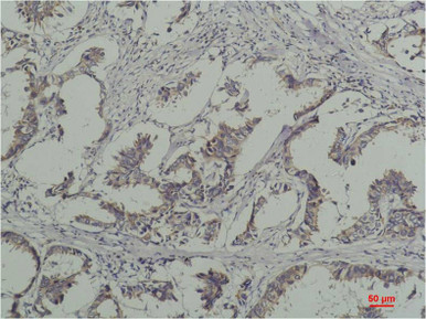
Anti-Phosphotyrosine Antibody
Detects phosphotyrosine — rare in the proteome but central to receptor and growth factor signalling.
View product →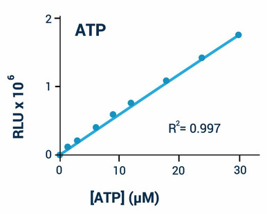
ATP Assay Kit (Luminescent)
Quantifies ATP directly, for work on energy status and mitochondrial function.
View product →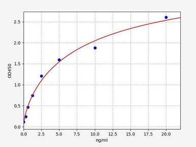
Human pAMPK ELISA Kit
Measures activated AMPK, the sensor that reads cellular energy state through the AMP to ATP ratio.
View product →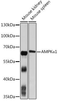
AMPK-alpha1 Rabbit Polyclonal Antibody
Total AMPK-alpha1, for normalising the phospho signal — the control that makes a phospho result interpretable.
View product →The phosphoryl group
Phosphorylation is the transfer of a phosphoryl group — a phosphorus atom with three oxygens — onto a substrate. The group carries two negative charges at physiological pH, and that is the source of its usefulness: attaching it introduces a large, highly charged, hydrogen-bonding moiety onto a protein surface, which alters local electrostatics and drives conformational change.
Two properties make phosphorylation the dominant regulatory modification in biology. It is reversible, because phosphatases remove what kinases add, so the modification behaves as a switch rather than a permanent mark. And it is fast, requiring no new protein synthesis, so a cell can respond within seconds and reverse the response just as quickly.
The same chemistry serves two quite different purposes. In metabolism, phosphoryl transfer stores and releases energy, principally through ATP. In signalling, it changes the activity of proteins. This article covers both, because the underlying reaction is identical even though the purposes are not.
The three types of phosphorylation
For ATP synthesis, three distinct mechanisms operate:
- Substrate-level phosphorylation — phosphate transferred directly from a high-energy substrate to ADP, in the cytosol and mitochondrial matrix.
- Oxidative phosphorylation — ATP synthesised indirectly, driven by a proton gradient generated by electron transport, in mitochondria.
- Photophosphorylation — the same indirect mechanism, but with light rather than substrate oxidation providing the energy, in chloroplasts.
The distinction that matters is direct versus indirect. Substrate-level phosphorylation is a single enzymatic transfer. The other two are chemiosmotic: energy is first stored as an electrochemical gradient, then converted to ATP by a separate enzyme. That separation is what allows a proton gradient to be used for other work, and what makes uncoupling possible.
Substrate-level phosphorylation
Here a phosphate group moves straight from a donor molecule to ADP, catalysed by a single enzyme. No membrane, no gradient, no oxygen — which is why this route keeps working under anaerobic conditions and is the only source of ATP in cells lacking mitochondria.
In glycolysis
Two steps of glycolysis generate ATP this way:
1,3-bisphosphoglycerate to 3-phosphoglycerate. Phosphoglycerate kinase transfers a phosphate from 1,3-BPG to ADP. The donor is an acyl phosphate with a high phosphoryl transfer potential, which is what makes the transfer favourable.
Phosphoenolpyruvate to pyruvate. Pyruvate kinase transfers phosphate from PEP to ADP. PEP has the highest phosphoryl transfer potential of any common metabolite, so this step is strongly exergonic and effectively irreversible.
Because glycolysis runs each step twice per glucose, these two reactions yield four ATP, against two consumed in the preparatory phase — a net gain of two ATP per glucose.
In the citric acid cycle
One step of the citric acid cycle also uses this mechanism: succinyl-CoA synthetase couples hydrolysis of the succinyl-CoA thioester bond to phosphorylation of GDP, producing GTP (or ATP directly, depending on the isoform and tissue). It is the only substrate-level phosphorylation in the cycle, with everything else generated later from the reduced electron carriers.
Oxidative phosphorylation
Oxidative phosphorylation generates most of the ATP in aerobic cells, and does so indirectly through two coupled stages.
Electron transport
NADH and FADH₂ produced by glycolysis, fatty acid oxidation and the citric acid cycle deliver electrons to a series of complexes in the inner mitochondrial membrane. Electrons pass down a chain of increasing reduction potential, ending at oxygen, which is reduced to water. The energy released at three of the complexes is used to pump protons from the matrix into the intermembrane space, generating an electrochemical gradient — the proton-motive force.
Oxygen’s role is specifically as the terminal electron acceptor. Without it, electrons have nowhere to go, the chain backs up, NADH accumulates and the whole process halts — which is why oxidative phosphorylation is strictly aerobic while substrate-level phosphorylation is not.
Chemiosmosis and ATP synthase
Protons flow back into the matrix through ATP synthase, and that flow drives ATP synthesis. The enzyme has two functional parts: a membrane-embedded proton channel that rotates as protons pass, and a catalytic head in which the rotation drives conformational changes that condense ADP and inorganic phosphate into ATP. It is a rotary molecular motor, and the coupling between proton flow and catalysis is mechanical rather than chemical.
Uncoupling
Because the two stages are separate, they can be uncoupled. If protons return across the membrane by another route, electron transport continues and oxygen is consumed, but no ATP is made — the energy dissipates as heat. Brown adipose tissue exploits this deliberately through uncoupling protein 1 to generate heat, and chemical uncouplers such as dinitrophenol do the same thing indiscriminately, which is why they are dangerous. Uncoupling is the clearest demonstration that the gradient, not a direct chemical intermediate, is what links oxidation to phosphorylation.
Oxidative versus substrate-level
| Feature | Oxidative phosphorylation | Substrate-level phosphorylation |
|---|---|---|
| Location | Inner mitochondrial membrane | Cytosol and mitochondrial matrix |
| Mechanism | Indirect — proton gradient drives ATP synthase | Direct transfer from a high-energy substrate to ADP |
| Energy source | Electrons from NADH and FADH₂ | Phosphoryl transfer potential of the substrate |
| Proton gradient | Required | Not involved |
| Oxygen | Required as terminal electron acceptor | Not required |
| ATP yield per glucose | Approximately 26 to 28 | 4 gross, 2 net from glycolysis, plus 2 GTP from the citric acid cycle |
| Occurs in | Aerobic respiration | Glycolysis and the citric acid cycle |
How much ATP does glucose actually yield?
Older textbooks give 36 to 38 ATP per glucose, and that figure still circulates widely. Current estimates are lower, at roughly 30 to 32 ATP, and the reason is worth understanding because it explains what the number represents.
The older calculation assumed whole-number stoichiometries — three ATP per NADH and two per FADH₂. Measurements show the proton-to-ATP ratio is not an integer: ATP synthase requires roughly four protons per ATP, while NADH oxidation pumps about ten and FADH₂ about six. That gives approximately 2.5 ATP per NADH and 1.5 per FADH₂.
There are also costs the older figure ignored. Importing cytosolic NADH into mitochondria consumes part of the gradient, and the shuttle used determines how much — the glycerol phosphate shuttle delivers electrons as FADH₂ rather than NADH, lowering the yield. Transporting ADP in and ATP out also draws on the proton-motive force.
So the honest answer is that the yield is a variable rather than a constant, depending on shuttle usage, tissue and how tightly coupled the mitochondria are. Around 30 to 32 is the usual figure quoted, and treating it as approximate is more accurate than treating any number as exact.
Photophosphorylation
Photophosphorylation generates ATP in plants, algae and cyanobacteria, using light instead of substrate oxidation as the energy source. It occurs in the thylakoid membrane of chloroplasts, and the machinery is closely analogous to the mitochondrial system — an electron transport chain, a proton gradient, and an ATP synthase.
Light absorbed by chlorophyll in the photosystems excites electrons, which pass along a carrier chain and pump protons into the thylakoid lumen. ATP synthase then converts that gradient into ATP.
Two modes operate, and the distinction matters. In non-cyclic photophosphorylation, electrons flow from water through photosystem II and photosystem I to NADP⁺, producing both ATP and NADPH and releasing oxygen. In cyclic photophosphorylation, electrons cycle back from photosystem I to the carrier chain, producing ATP only, with no NADPH and no oxygen release. Plants use the cyclic route to adjust the ATP to NADPH ratio when carbon fixation demands more ATP than the linear pathway supplies.
Protein phosphorylation as a switch
Beyond energy metabolism, phosphorylation is the principal mechanism by which protein activity is regulated. Kinases add phosphate; phosphatases remove it; the steady state reflects the balance between them.
| Residue | Relative abundance | Typical context |
|---|---|---|
| Serine | By far the most common, roughly 85 to 90% of sites | Broad regulatory phosphorylation by serine/threonine kinases such as PKA, PKC, CDKs and AKT |
| Threonine | Around 10 to 15% of sites | Often adjacent to serine sites; activation-loop phosphorylation in many kinases |
| Tyrosine | Under 2% of sites | Receptor and growth factor signalling; creates docking sites for SH2 domains |
Tyrosine phosphorylation is worth singling out precisely because it is rare. Its scarcity makes it a low-noise signal, and phosphotyrosine residues serve as specific docking sites recognised by SH2 and PTB domains — so the modification does not merely change conformation, it creates a binding site that recruits new partners.
What phosphorylation actually does to a protein
- Changes conformation and activity. Adding charge in or near an active site can activate or inhibit an enzyme; activation-loop phosphorylation is how most kinases are themselves switched on.
- Creates or destroys binding sites. Phosphotyrosine recruits SH2 domains; phosphoserine and phosphothreonine recruit 14-3-3 proteins and others.
- Controls localisation. Phosphorylation can mask or expose nuclear import and export signals, moving a protein between compartments.
- Controls stability. Phosphorylation frequently marks a protein for ubiquitination and proteasomal degradation, setting its half-life.
A brief note on breadth: in bacteria and plants, two-component systems phosphorylate histidine and aspartate residues rather than serine, threonine or tyrosine. Those sites are chemically labile and largely invisible to standard mammalian phospho-reagents, which is why they are under-represented in the literature relative to their biological importance.
Measuring phosphorylation
The central experimental point is that phosphorylation changes a protein’s state, not its amount. Total protein barely moves on stimulation, so any approach that measures abundance will report nothing.
- Phospho-specific antibodies are the workhorse. Site-specific antibodies target one residue; pan-specific antibodies against phosphoserine, phosphothreonine or phosphotyrosine survey the whole proteome and are useful when the relevant site is unknown.
- Always run the total protein alongside. A rise in phospho signal means nothing unless total protein is unchanged — otherwise expression, not phosphorylation, may have changed.
- Include phosphatase inhibitors in every lysis buffer. Cellular phosphatases remain active in lysate and strip phosphate within minutes, and their omission is the commonest cause of a falsely negative phospho blot.
- Take a time course. Phosphorylation typically peaks within minutes to an hour and resolves as phosphatases act, so a single late timepoint can miss the event entirely.
- Phosphoproteomics by mass spectrometry with phosphopeptide enrichment identifies sites without prior knowledge, and gives stoichiometry that antibodies cannot.
- Functional readouts close the loop — ATP concentration for energy metabolism, or a downstream substrate for signalling, confirms that the phosphorylation had a consequence.
Choosing phospho reagents
Pan-specific antibodies against phosphoserine, phosphothreonine and phosphotyrosine, site-specific phospho antibodies, ATP assays and matched phospho and total pairs for normalisation.
Browse phospho antibodies & assays →Frequently asked questions
What are the three types of phosphorylation?
Substrate-level phosphorylation, which transfers phosphate directly from a high-energy substrate to ADP; oxidative phosphorylation, which uses a proton gradient generated by electron transport; and photophosphorylation, which does the same but powered by light in chloroplasts.
How much ATP does one glucose molecule produce?
Around 30 to 32 by current estimates, not the 36 to 38 given in older textbooks. The revision reflects non-integral proton-to-ATP stoichiometry — roughly 2.5 ATP per NADH and 1.5 per FADH₂ — plus the energetic cost of transporting NADH, ADP and ATP across the membrane.
What is the difference between substrate-level and oxidative phosphorylation?
Substrate-level is direct: one enzyme moves phosphate from a substrate to ADP, needing no oxygen or membrane. Oxidative is indirect: electron transport builds a proton gradient and ATP synthase converts that gradient into ATP, and it requires oxygen as the terminal electron acceptor.
Which amino acids are phosphorylated?
In eukaryotes, principally serine (roughly 85 to 90% of sites), threonine (10 to 15%) and tyrosine (under 2%). Bacteria and plants also phosphorylate histidine and aspartate in two-component signalling systems.
Why is tyrosine phosphorylation important if it is so rare?
Because scarcity makes it a low-noise signal, and phosphotyrosine acts as a specific docking site for SH2 and PTB domains. It therefore recruits new binding partners rather than merely altering conformation, which is why receptor signalling relies on it.
What does uncoupling mean?
Allowing protons to cross the inner mitochondrial membrane without passing through ATP synthase. Electron transport and oxygen consumption continue but no ATP is made, and the energy is released as heat. Brown fat does this deliberately via uncoupling protein 1.
Why do I need to measure total protein as well as phospho?
Because phosphorylation changes state, not abundance. Without the total, an increased phospho signal cannot be distinguished from increased expression of the protein. Phospho over total is the meaningful quantity.
Rithika Suresh completed her undergraduate degree in Biotechnology in Anna University before completing her masters in Biotechnology at University College Dublin.
Recent Posts
-
Suspected Plague Death at a Siberian Anti-Plague Institute: What Researchers Should Know
Infectious Disease · News AnalysisSuspected Plague Death at a Siberian Anti-Plague Institute: What …6th Oct 2026 -
LPS ELISA Troubleshooting: The Importance of Sample Nature
Quick answer A researcher measuring circulating lipopolysaccharide (LPS) in human serum …25th Sep 2026 -

Sekundärantikörper wählen: Die Regel, die im Labor wirklich zählt
Sekundärantikörper wählen: Die Regel, die im Labor wirklich zählt Wählen Sie einen Sekundärantikörp …24th Sep 2026