
Protein Kinases: Overview, Classification and Therapeutic Potential
Protein Kinases: Function, Classification, Structure and Inhibitors
Protein kinases transfer a phosphate group from ATP onto specific residues of target proteins, switching their activity on or off. Around 500 of them make up the human kinome, and because they sit at the control points of nearly every pathway, they are among the most heavily pursued drug targets in medicine. This guide covers what they do, how they are classified, their conserved architecture, how they are regulated, and how inhibitors work.
Browse kinase proteins →Key takeaways
- Kinases catalyse transfer of the gamma-phosphate of ATP directly onto a substrate hydroxyl group, leaving ADP — ATP is not first broken down to free phosphate.
- Three classes are defined by target residue: serine/threonine kinases, tyrosine kinases, and dual-specificity kinases that act on both.
- Tyrosine kinases divide into receptor tyrosine kinases, which are transmembrane proteins, and non-receptor tyrosine kinases, which are cytoplasmic.
- All eukaryotic protein kinases share a bilobal catalytic domain, with ATP bound in the cleft between the lobes and an activation loop whose phosphorylation usually switches the enzyme on.
- Regulation operates through phosphorylation, allosteric binding, subcellular localisation, scaffolding and feedback — rarely through one mechanism alone.
- Dysregulated kinase activity underlies cancers, inflammatory and autoimmune disease, metabolic disorders and several developmental syndromes.
- Most approved kinase inhibitors are ATP-competitive small molecules; resistance and off-target activity remain the central challenges.
Kinase proteins and assay reagents
The panel below spans the three classes described in this guide — a serine/threonine kinase, receptor and non-receptor tyrosine kinases, and a dual-specificity kinase — plus a phospho-specific antibody for reading activity downstream.
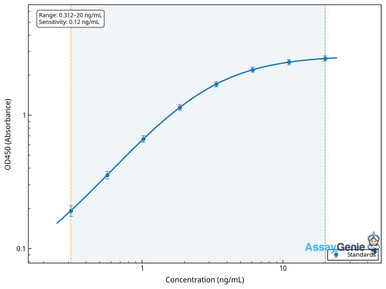
Human MAP3K5 / ASK1 ELISA Kit
A serine/threonine kinase central to stress-activated signalling upstream of JNK and p38.
View product →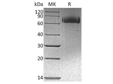
Human EGFR / ErbB1 Recombinant Protein
The canonical receptor tyrosine kinase, and the target of the first generation of targeted cancer drugs.
View product →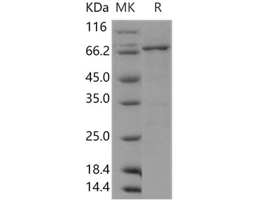
Human DDR1 Kinase Recombinant Protein (Active)
A collagen-activated receptor tyrosine kinase, supplied active for in vitro kinase assays.
View product →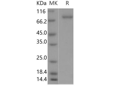
Human SRC / c-SRC Recombinant Protein (Active)
The archetypal non-receptor tyrosine kinase — cytoplasmic, membrane-anchored, and active for kinase assays.
View product →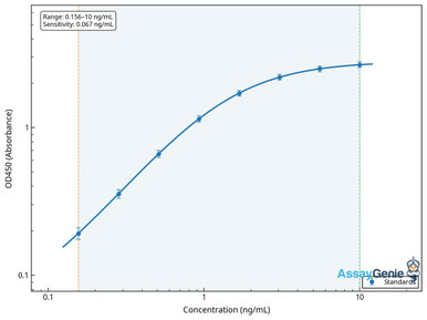
Human MEK1 / MAP2K1 ELISA Kit
A dual-specificity kinase and the MEK in the RAF-MEK-ERK cascade targeted by MEK inhibitors.
View product →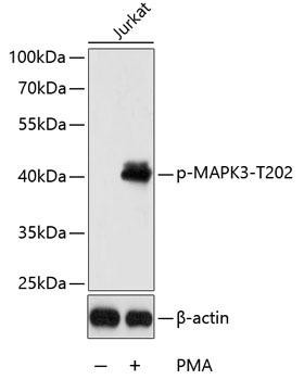
Phospho-ERK1 (Thr202) Rabbit Polyclonal Antibody
Detects activated ERK1 — the standard downstream readout of MAPK pathway signalling.
View product →What are protein kinases?
Protein kinases are enzymes that attach a phosphate group to specific amino acid residues on target proteins. Adding a bulky, doubly negative phosphate to a serine, threonine or tyrosine side chain alters the local charge and shape of the protein, which in turn changes its conformation, its activity, its binding partners or its stability. Because phosphatases remove those phosphates, the modification is reversible — and reversibility is what makes it useful as a switch rather than a one-way commitment.
Around 500 protein kinases are encoded in the human genome, collectively the kinome, making them one of the largest enzyme families. They control cell growth and division, differentiation, metabolism, apoptosis, immune activation and essentially every signal transduction pathway. That central position is why kinase dysregulation causes so much disease, and why the family accounts for a large share of modern targeted therapeutics.
The catalytic mechanism
The chemistry is worth stating precisely, because it is often described incorrectly. ATP binds in the cleft of the kinase catalytic domain together with a magnesium ion, which coordinates the phosphate groups and positions them for reaction. The hydroxyl group of the substrate residue — serine, threonine or tyrosine — then attacks the gamma-phosphate of ATP directly, in a single in-line phosphoryl transfer. The products are the phosphorylated substrate and ADP.
ATP is not first hydrolysed to ADP and free inorganic phosphate, with that free phosphate subsequently attacking the substrate. That description appears frequently and is mechanistically wrong: free phosphate in solution is not a competent phosphorylating agent, and the reaction is a direct transfer within the enzyme active site. The distinction matters practically as well, because it explains why kinases require magnesium and why the great majority of inhibitors work by occupying the ATP site.
Classification
The primary division is by which residue the enzyme phosphorylates.
| Class | Target residues | Location and character | Examples |
|---|---|---|---|
| Serine/threonine kinases (STKs) | Serine and threonine | Mostly cytoplasmic and nuclear; the largest group | PKA, PKC, CDKs, AKT, GSK-3, mTOR, RAF |
| Receptor tyrosine kinases (RTKs) | Tyrosine | Transmembrane — extracellular ligand-binding domain, single-pass helix, intracellular kinase domain | EGFR, insulin receptor, VEGFR, FGFR, RET, DDR1 |
| Non-receptor tyrosine kinases | Tyrosine | Cytoplasmic, often membrane-anchored or associated with receptors | SRC, ABL, JAK, FAK, SYK, BTK |
| Dual-specificity kinases (DSKs) | Both serine/threonine and tyrosine | Cytoplasmic and nuclear | MEK1/2 (MAP2K), DYRKs, WEE1 |
Two points in the tyrosine kinase row are commonly reversed and worth being explicit about. Receptor tyrosine kinases are transmembrane proteins, not cytoplasmic — that is precisely what makes them receptors, with the ligand-binding domain outside the cell and the catalytic domain inside. Ligand binding drives dimerisation and autophosphorylation of the intracellular domain, creating docking sites for downstream adaptors. Non-receptor tyrosine kinases are the cytoplasmic ones, and they are not defined by nuclear localisation or DNA binding. Some, such as ABL, do shuttle to the nucleus under particular conditions, but that is a feature of individual kinases rather than a property of the class.
Kinases are also grouped functionally — signalling, metabolic and housekeeping — and structurally into groups such as AGC, CAMK, CK1, CMGC, STE and TK, which is the classification used by kinome-wide studies and selectivity profiling.
Structure of the catalytic domain
Every eukaryotic protein kinase shares a conserved catalytic domain of roughly 250 to 300 residues, folded into two lobes with ATP bound in the cleft between them.
- The N-terminal lobe is smaller and mostly beta-sheet, carrying the glycine-rich loop that positions the ATP phosphates, and a conserved lysine and glutamate pair whose salt bridge marks the active conformation.
- The C-terminal lobe is larger and mostly helical, and provides the substrate-binding surface and the catalytic residues.
- The activation loop sits in the C-lobe. In most kinases it must itself be phosphorylated before the enzyme becomes fully active, which is how kinase cascades propagate.
- The hinge and gatekeeper residue connect the lobes and line the back of the ATP pocket. Differences here between kinases are a major source of inhibitor selectivity, and mutation of the gatekeeper is a classic resistance mechanism.
Beyond this conserved core, kinases differ enormously. Regulatory regions vary from autoinhibitory pseudosubstrate segments to SH2 and SH3 domains, pleckstrin homology domains, and cyclin-binding surfaces. It is therefore misleading to describe a universal architecture of catalytic, regulatory and effector domains — the catalytic domain is conserved, but what flanks it is specific to each family and is often exactly what determines how that kinase is switched on.
Kinases in signal transduction
Kinases convert extracellular information into intracellular action, and they do it in cascades. A ligand binds a receptor, the receptor kinase autophosphorylates, adaptors dock, and a series of kinases phosphorylate one another in sequence until effectors are reached — transcription factors, metabolic enzymes, cytoskeletal proteins.
The cascade architecture is not redundancy. It amplifies, since each active kinase phosphorylates many substrates; it integrates, since each tier can receive inputs from several pathways; and it provides multiple points for regulation and for pharmacological intervention. The RAF-MEK-ERK module is the textbook case, and it also illustrates why inhibiting one tier often produces resistance through reactivation at another.
Phosphorylation suits this role because it is fast, reversible and does not require new protein synthesis. A cell can respond within seconds and reverse the response just as quickly once phosphatases act.
How kinases are regulated
Unrestrained kinase activity is dangerous, so kinases are controlled by several overlapping mechanisms rather than any single one.
- Activation-loop phosphorylation. The commonest switch, and how one kinase activates the next.
- Autoinhibition. Many kinases carry segments that occlude their own active site until displaced by a ligand or a modification.
- Allosteric regulation. Binding of second messengers such as cyclic AMP or calcium, or of partner proteins such as cyclins, shifts the enzyme between inactive and active conformations.
- Cofactor and substrate availability. Activity depends on magnesium and on ATP concentration, which is one route by which metabolic state feeds into signalling.
- Localisation and scaffolding. Scaffold proteins hold kinases with their substrates in defined compartments, so the same kinase can produce different outcomes depending on where it is held.
- Opposition by phosphatases. Steady-state phosphorylation reflects the balance between kinase and phosphatase, so a change in phosphatase activity alters signalling without any change in the kinase.
- Degradation and feedback. Ubiquitin-mediated turnover and negative feedback loops set the duration of a response.
Disease associations
Because kinases sit at pathway control points, mutations that lock them on — or overexpression that raises their output — have disproportionate consequences.
| Disease area | Kinases implicated | How dysregulation contributes |
|---|---|---|
| Cancer | BCR-ABL, EGFR, BRAF, ALK, HER2, RET | Constitutive activation from fusion, mutation or amplification drives proliferation and survival independently of normal signals |
| Neurodegeneration | GSK-3β, CDK5, LRRK2 | Aberrant activity promotes pathological protein phosphorylation and aggregation, notably of tau |
| Cardiovascular disease | ERK1/2, JNK, PKC, AKT | Contributes to cardiac hypertrophy, and to ischaemia-reperfusion injury and heart failure |
| Inflammatory and autoimmune disease | JAK1/2/3, SYK, BTK | Sustains immune cell activation and cytokine production in rheumatoid arthritis, psoriasis and inflammatory bowel disease |
| Metabolic disease | AMPK, insulin receptor kinase | Impaired signalling disrupts glucose and lipid handling in insulin resistance and type 2 diabetes |
| Developmental syndromes | MEK1/2, RAF, FGFR | Germline activating mutations cause Noonan, Costello, Apert and craniosynostosis syndromes |
One correction to a claim that circulates in this context: c-Myc is not a protein kinase. It is a basic helix-loop-helix transcription factor, and although MYC dysregulation is genuinely one of the commonest events in cancer, it acts by driving transcription rather than by phosphorylating substrates. Kinases do sit both upstream and downstream of MYC — which is likely the source of the confusion — but it does not belong in a list of serine/threonine kinases.
Kinase inhibitors
Kinase inhibitors are among the most successful classes of targeted therapy. They are distinct from classical cytotoxic chemotherapy: rather than damaging dividing cells indiscriminately, they block a specific enzyme that a tumour or an inflammatory process depends on.
How they work
Most approved inhibitors are small molecules that occupy the ATP-binding cleft, competing with ATP and preventing phosphoryl transfer. Several mechanistic classes are distinguished:
- Type I bind the ATP site of the active conformation — for example gefitinib against EGFR.
- Type II bind the ATP site of the inactive conformation, reaching into an adjacent pocket, which often improves selectivity — imatinib against BCR-ABL is the classic example.
- Type III and IV bind allosteric sites away from the ATP cleft, so they are not competing with high intracellular ATP concentrations. Several MEK inhibitors work this way.
- Covalent inhibitors form an irreversible bond with a cysteine in the active site — ibrutinib against BTK, and osimertinib against mutant EGFR.
Approved examples
Imatinib transformed chronic myeloid leukaemia by inhibiting the BCR-ABL fusion kinase. Gefitinib, erlotinib and osimertinib target EGFR in non-small cell lung cancer. BRAF inhibitors combined with MEK inhibitors are standard in BRAF-mutant melanoma. Outside oncology, JAK inhibitors are approved for rheumatoid arthritis and other inflammatory conditions, and BTK inhibitors for B-cell malignancies.
Two clarifications, since both errors appear in secondary sources. Gemcitabine is not a kinase inhibitor — it is a nucleoside analogue antimetabolite that interferes with DNA synthesis, and it does not target PDK1. And AP20187 is not a kinase inhibitor either — it is a bivalent chemical dimeriser used to induce protein dimerisation in engineered systems, a laboratory tool rather than a therapeutic.
Remaining challenges
Selectivity is intrinsically difficult, because the ATP site is conserved across a family of roughly 500 enzymes, so off-target activity is common and sometimes therapeutically useful. Resistance emerges readily, through gatekeeper mutations that block inhibitor binding, amplification of the target, or reactivation of the pathway at another tier. And because many kinases are required in normal tissue, the therapeutic window can be narrow. Selectivity profiling across the kinome and rational combination strategies are the main responses to these problems.
Choosing kinase reagents
Recombinant kinases including active enzymes for in vitro assays, ELISA kits for kinase quantification, and phospho-specific antibodies for reading pathway activity.
Browse kinase reagents →Frequently asked questions
What does a protein kinase actually do?
It transfers the gamma-phosphate of ATP onto a serine, threonine or tyrosine residue of a target protein. The substrate hydroxyl attacks the ATP phosphate directly in the active site, producing the phosphorylated protein and ADP. ATP is not first broken down into ADP and free phosphate.
What are the three classes of protein kinase?
Serine/threonine kinases, tyrosine kinases and dual-specificity kinases, defined by which residues they phosphorylate. Tyrosine kinases divide further into transmembrane receptor tyrosine kinases and cytoplasmic non-receptor tyrosine kinases.
Are receptor tyrosine kinases cytoplasmic?
No — they are transmembrane, with an extracellular ligand-binding domain and an intracellular kinase domain. The cytoplasmic tyrosine kinases are the non-receptor class, such as SRC, ABL and JAK.
Why do kinases need magnesium?
Magnesium ions coordinate the phosphate groups of ATP within the active site, positioning them correctly for transfer and stabilising the transition state. Without it, catalysis is not efficient, which is why kinase assay buffers always include magnesium.
Why is kinase inhibitor selectivity so difficult?
Because most inhibitors bind the ATP pocket, and that pocket is conserved across roughly 500 human kinases. Achieving selectivity means exploiting subtle differences at the hinge and gatekeeper region, or targeting allosteric sites outside the ATP cleft.
Is c-Myc a kinase?
No. c-Myc is a basic helix-loop-helix transcription factor. Its dysregulation is a very common event in cancer, but it acts through transcription rather than phosphorylation, and it is sometimes incorrectly listed among serine/threonine kinases.
What is a gatekeeper mutation?
A change to the residue lining the back of the ATP pocket that prevents an inhibitor from binding while leaving the kinase active. It is one of the principal routes to acquired resistance against ATP-competitive inhibitors.
Rithika Suresh completed her undergraduate degree in Biotechnology in Anna University before completing her masters in Biotechnology at University College Dublin.
Recent Posts
-
Suspected Plague Death at a Siberian Anti-Plague Institute: What Researchers Should Know
Infectious Disease · News AnalysisSuspected Plague Death at a Siberian Anti-Plague Institute: What …6th Oct 2026 -
LPS ELISA Troubleshooting: The Importance of Sample Nature
Quick answer A researcher measuring circulating lipopolysaccharide (LPS) in human serum …25th Sep 2026 -

Sekundärantikörper wählen: Die Regel, die im Labor wirklich zählt
Sekundärantikörper wählen: Die Regel, die im Labor wirklich zählt Wählen Sie einen Sekundärantikörp …24th Sep 2026