
p38 MAPK Signaling Review
The p38 MAPK signaling pathway is an intracellular signaling pathway that plays a crucial role in cellular responses to various stressors and inflammatory stimuli. It is a part of the larger MAPK superfamily, which also includes ERK and JNK pathways. The p38 MAPK pathway is involved in regulating a wide range of cellular processes, including cell proliferation, differentiation, apoptosis, inflammation, and immune responses.
Key Takeaways:
- p38 MAPK, part of the MAPK family, is key in cellular responses to stress and inflammation.
- This pathway regulates cell processes like proliferation, differentiation, apoptosis, and immune responses.
- The MAPK family includes ERKs, JNKs, and p38 MAPKs, each responding to different stimuli.
- p38 MAPK has four isoforms, each with specific roles in various cellular functions.
- It interacts with ERK and JNK pathways, highlighting its role in complex cellular regulation.
- p38 MAPK's involvement in tumor suppression and response to cellular stress underscores its significance in cancer biology.
MAPK Family
The MAPK family is a group of serine/threonine protein kinases that play essential roles in transducing extracellular signals into a wide range of cellular responses. These kinases are highly conserved across species and are found in various eukaryotic organisms. The three main subfamilies of MAPKs are:
-
ERKs (Extracellular Signal-Regulated Kinases) are primarily activated by growth factors and mitogens. They are involved in regulating cell proliferation, differentiation, survival, and other processes related to cell growth and development.
-
JNKs (c-Jun N-terminal Kinases) are activated in response to environmental stresses, cytokines, and inflammatory signals. They play critical roles in apoptosis, immune responses, and cellular responses to stress.
-
p38 MAPKs are activated by various stress stimuli, inflammatory cytokines, and cellular damage. They are key regulators of inflammation, immune responses, stress responses, and cell fate decisions.
MAPKKK (MAPK Kinase Kinase) Genes
MAPKKKs are upstream kinases that activate MAPKKs, initiating the MAPK signaling cascade. Several important MAPKKKs include:
-
MEKK1 (MAP3K1) activates both the JNK and p38 MAPK pathways in response to stress and inflammatory signals.
-
MEKK4 (MAP3K4) also activates the JNK and p38 MAPK pathways, influencing cell proliferation, apoptosis, and immune responses.
-
ASK1 (MAP3K5) specifically activates the JNK and p38 MAPK pathways in response to oxidative stress and cytokines, playing a role in cell survival and apoptosis.
- TAK1 (MAP3K7) is a versatile MAPKKK that can activate multiple MAPK pathways, including p38, JNK, and NF-κB signaling. It is an essential regulator of inflammatory responses and immune signaling.
These MAPKKKs are part of the initial tier in the MAPK signaling cascade. They phosphorylate and activate specific MAPKKs, which, in turn, phosphorylate and activate their corresponding downstream MAPKs. The activated MAPKs then modulate the activity of various downstream effectors, leading to diverse cellular responses.
p38 MAPK Family
The p38 MAPK family exhibits remarkable diversity, with several isoforms and distinct roles in various cellular processes. Four isoforms have been identified: p38α (MAPK14), p38β (MAPK11), p38γ (MAPK12), and p38δ (MAPK13). Each isoform shares a conserved structure, containing a kinase domain and regulatory regions, but they also display unique expression patterns, activation mechanisms, and substrate specificities.
p38α (MAPK14): p38α is the most extensively studied isoform and is ubiquitously expressed in many tissues. It plays a crucial role in mediating cellular responses to stress, inflammatory cytokines, and various environmental stimuli. p38α regulates diverse processes, including cell cycle progression, apoptosis, immune responses, and cell differentiation.

| Human Mitogen Activated Protein Kinase 14 (MAPK14) ELISA Kit | |
|---|---|
| ELISA Type | Sandwich |
| Sensitivity | 0.56ng/mL |
| Range | 1.56-100ng/mL |
p38β (MAPK11): p38β exhibits a more restricted expression pattern and is predominantly found in skeletal muscle and heart tissues. Its specific functions and roles in cellular responses are less well-characterized compared to p38α, but it is believed to be involved in muscle differentiation and inflammation.
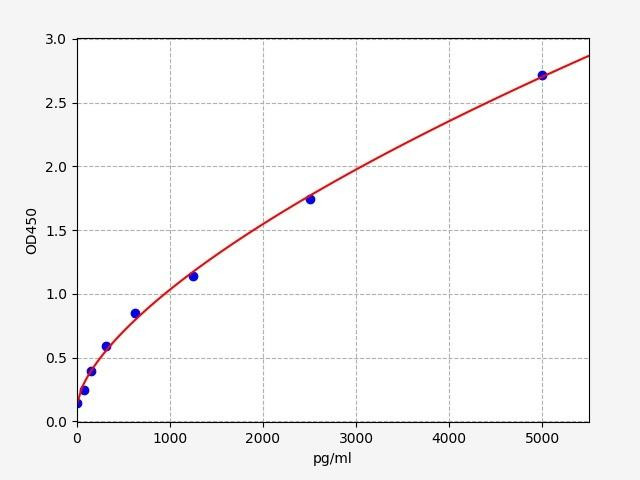
| Human MAPK11 (Mitogen-activated protein kinase 11) ELISA Kit (HUFI03437) | |
|---|---|
| ELISA Type | Sandwich |
| Sensitivity | 46.875pg/ml |
| Range | 78.125-5000pg/ml |
p38γ (MAPK12): p38γ is expressed in various tissues, including the brain, heart, and immune cells. It is activated by different stimuli than p38α and has distinct substrate specificity. p38γ is implicated in regulating cell migration, cell survival, and immune responses.

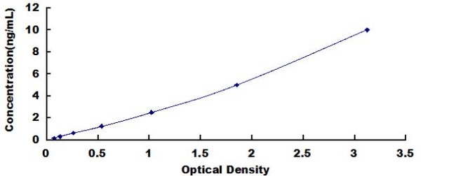
| Human Mitogen Activated Protein Kinase 12 (MAPK12) ELISA Kit | |
|---|---|
| ELISA Type | Sandwich |
| Sensitivity | 0.072ng/mL |
| Range | 0.156-10ng/mL |
p38δ (MAPK13): p38δ is expressed in various tissues, including the brain, liver, and adipose tissue. It is responsive to specific stress and growth factors and has been associated with adipogenesis and insulin sensitivity.
| Human Mitogen Activated Protein Kinase 13 (MAPK13) ELISA Kit | |
|---|---|
| ELISA Type | Sandwich |
| Sensitivity | 0.061ng/mL |
| Range | 0.156-10ng/mL |
p38 MAPK Signaling
The p38 MAPK pathway is a highly conserved intracellular signaling cascade that plays a central role in cellular responses to various stressors and inflammatory stimuli. The p38 MAPK pathway is activated by diverse extracellular signals, including pro-inflammatory cytokines (e.g., TNF-alpha, IL-1β), environmental stresses (e.g., heat shock, osmotic stress), and cellular damage.
p38 Pathway
1. p38 MAPK Activation
p38 MAPK activation is a crucial step in its signaling cascade, governing cellular responses to various stress stimuli. Full activation of p38 MAPK requires the dual phosphorylation of Thr and Tyr residues on the activation loop, specifically within the Thr-Gly-Tyr motif located on the kinase subdomain VIII. This phosphorylation induces conformational changes, facilitating substrate recognition and increasing the kinase's activity.
The primary MAP2Ks responsible for the phosphorylation of p38 MAPK are MKK3 and MKK6. Upon stress stimulation, these MAP2Ks phosphorylate Thr and Tyr residues, leading to p38 MAPK activation. Moreover, p38α isoform can be activated through noncanonical mechanisms. For instance, T cell receptor (TCR) stimulation induces Tyr 323 phosphorylation by the kinases ZAP70 and p56lck, promoting p38α autophosphorylation and subsequent activation. Binding to the protein TAB1 also leads to autophosphorylation and activation of p38α.
Additionally, a third noncanonical and MAP2K-independent mechanism leading to p38 signaling has been observed in tumor cells depleted of the DNA replication initiation factor Cdc7. Under replication stress, these cells undergo programmed cell death in an ATR- and p38-dependent manner, suggesting that p38 mediates apoptosis upon replication stress.
Once activated, p38 MAPK translocates to the nucleus or other cellular compartments, where it phosphorylates various downstream targets, thereby initiating specific cellular responses. This process involves the modulation of several transcription factors, protein kinases, and other signaling molecules that regulate critical cellular processes, including inflammation, cell differentiation, apoptosis, and stress responses.
2. p38 in Upstream Kinases (MKK3 and MKK6)
Two primary MAPK Kinases (MAPKKs) that activate p38 MAPK are MAPK Kinase 3 (MKK3) and MAPK Kinase 6 (MKK6). In response to cellular stress, specific MAPK Kinase Kinases (MAPKKKs), such as ASK1 (Apoptosis Signal-regulating Kinase 1) or MEKK1 (Mitogen-Activated Protein Kinase Kinase Kinase 1), become activated and initiate the phosphorylation cascade leading to the activation of MKK3 and MKK6.
MKK3 and MKK6, once activated, serve as dedicated kinases that directly target and phosphorylate specific threonine and tyrosine residues within the activation loop of p38 MAPK. The dual phosphorylation of p38 MAPK leads to its full activation, transforming it into an active kinase.
3. Downstream Signaling of p38 MAPK
The fully activated p38 MAPK, phosphorylates several key downstream targets, including various transcription factors, such as ATF-2 (Activating Transcription Factor 2), Elk-1 (Ets-Like Protein 1), and CHOP (C/EBP Homologous Protein). These transcription factors modulate the expression of specific genes involved in the cellular response to stress and inflammation.
p38 Phosphorylation Targets
Activated p38 MAPK phosphorylates a wide array of substrates, including transcription factors, protein kinases, and other regulatory proteins. Some important phosphorylation targets of p38 MAPK include:
- Transcription Factors:
-
- ATF-2 (Activating Transcription Factor 2): Phosphorylation of ATF-2 by p38 MAPK enhances its transcriptional activity, leading to the upregulation of target genes involved in stress responses and inflammation.
- c-Jun: p38-mediated phosphorylation of c-Jun enhances its dimerization with c-Fos, forming the AP-1 transcription factor, which regulates genes associated with proliferation, apoptosis, and immune responses.
-
- Other Protein Kinases:
-
- MAPKAPK-2 (MAPK-Activated Protein Kinase-2): p38 MAPK activates MAPKAPK-2 by phosphorylation, leading to the phosphorylation of downstream targets, such as heat shock protein HSP27, involved in cytoskeletal rearrangement and cell migration.
- MSK1/2 (Mitogen- and Stress-Activated Protein Kinase 1/2): Phosphorylation of MSK1/2 by p38 MAPK contributes to the activation of specific transcription factors involved in inflammation and immune responses.
-
- Regulatory Proteins:
-
- MK2 (MAPK-Activated Protein Kinase 2): p38 MAPK phosphorylates and activates MK2, which in turn modulates mRNA stability and translation, affecting the expression of genes involved in inflammation and cell cycle regulation.
- HSP27 (Heat Shock Protein 27): Phosphorylation of HSP27 by p38 MAPK facilitates its chaperone activity, protecting cells from stress-induced damage.
-
- Other Downstream Targets:
-
- p38 MAPK also phosphorylates various cytoskeletal and signaling proteins, ion channels, and other factors that contribute to specific cellular responses, including cell migration, adhesion, and survival.
-
The phosphorylation of these targets orchestrates the diverse cellular responses mediated by the p38 MAPK pathway, allowing cells to adapt to changes in their environment and appropriately respond to stress and inflammatory stimuli.
Regulation of p38 MAPK Signaling
The p38 MAPK signaling pathway is tightly regulated to ensure appropriate cellular responses and prevent excessive activation. Specific protein phosphatases, such as MKP-1 (MAPK phosphatase-1), dephosphorylate and inactivate p38 MAPK, providing negative feedback regulation. Additionally, MAPKKK inhibitors, like suppressor of cytokine signaling (SOCS) and TRAF-interacting protein (TRIP), act as negative regulators by inhibiting MAPKKKs. Scaffolding proteins, such as JIP (JNK-interacting protein), can sequester components of the p38 MAPK pathway, restricting their interactions and activity. Moreover, the p38 MAPK pathway interacts with other signaling pathways, including the ERK and JNK pathways, through cross-phosphorylation and regulatory feedback loops, which fine-tune cellular responses.
p38 Functions and Cellular Responses
The p38 MAPK pathway plays a pivotal role in a wide range of cellular responses. It regulates the production of pro-inflammatory cytokines (e.g., TNF-alpha, IL-1β, IL-6) and chemokines, contributing to immune responses and inflammation. Additionally, it is involved in the activation of immune cells like macrophages and dendritic cells. In response to stressors such as oxidative stress, heat shock, osmotic stress, and DNA damage, p38 MAPK helps cells adapt by mediating stress-induced apoptosis or cell cycle arrest. It influences the cell cycle by modulating the activities of cyclins and cyclin-dependent kinases (CDKs), either promoting cell cycle arrest or progression, depending on the cellular context. The p38 MAPK pathway participates in regulating cell differentiation processes, such as osteoblast and adipocyte differentiation. It can also mediate either pro-apoptotic or pro-survival effects, depending on the cell type and context.
p38 MAPK as a Tumor Suppressor
p38 MAPK, in addition to its well-known roles in stress response and inflammation, has also been recognized as a tumor suppressor in certain contexts. As a member of the mitogen-activated protein kinase (MAPK) family, p38 plays a crucial role in regulating cell growth, differentiation, and apoptosis. Dysregulation of these processes can contribute to the development and progression of cancer.
Several lines of evidence support the tumor-suppressive functions of p38 MAPK. Activation of p38 has been shown to induce cell cycle arrest and inhibit cell proliferation, preventing the uncontrolled growth of cancer cells. Additionally, p38 activation can trigger apoptosis in response to various stress signals, providing a mechanism for eliminating damaged or potentially malignant cells.
Furthermore, p38 MAPK is involved in the regulation of the epithelial-mesenchymal transition (EMT), a process that influences cell migration and invasion during metastasis. Activation of p38 has been found to suppress EMT, thus reducing the ability of cancer cells to spread and invade surrounding tissues.
Moreover, p38 MAPK can modulate the tumor microenvironment by regulating the production of cytokines, chemokines, and growth factors. This can influence immune responses and angiogenesis, potentially affecting tumor growth and metastasis.
p38 MAPK Regulation of Cell Death
The p38 MAPK pathway plays a pivotal role in controlling cell fate, particularly in orchestrating cellular responses to stress-induced cell death. Upon treatment of cells with sodium arsenite, p38 MAPK is activated, and its pivotal role in cellular responses to stress becomes apparent. One key aspect of this regulation involves the activation of the forkhead transcription factor, FOXO3a. Activated p38 MAPK triggers the activation of FOXO3a, which in turn upregulates the pro-apoptotic protein BimEL. Furthermore, p38 MAPK has been shown to directly phosphorylate BimEL in response to arsenite treatment, resulting in cell death. These findings highlight the role of p38 MAPK in regulating the pro-apoptotic protein Bim, and its significance in cellular responses to stress-induced cell death.
p38 MAPK and Interactions with Ras and JNK
The p38 MAPK signaling pathway does not act in isolation but rather interacts with other crucial signaling pathways, such as the extracellular signal-regulated kinases 1 and 2 (ERK1/2) and the c-Jun N-terminal Kinases (JNK), along with Ras GTPase activation, to fine-tune cellular responses to various stimuli.
Emerging evidence suggests extensive crosstalk and functional connections between ERK1/2, JNK, and p38 MAPK pathways. These interactions can occur at multiple levels, including feedback loops, shared upstream activators, and coordinated regulation of downstream targets. For instance, while p38 MAPK and JNK pathways often collaborate to promote cellular apoptosis in response to certain stressors, ERK1/2 signaling may exert anti-apoptotic effects to counterbalance these responses. Moreover, various cellular stimuli can differentially regulate the activation of these MAPK pathways, leading to distinct cellular outcomes or even influencing cell fate decisions.
ERK1/2 Pathway
The ERK1/2 pathway, also known as the Ras-Raf-MEK-ERK pathway, plays a pivotal role in mediating cell proliferation, differentiation, and survival in response to growth factors and mitogenic signals. Activation of the pathway involves the small GTPase Ras, which triggers a phosphorylation cascade, leading to the activation of the serine/threonine kinases Raf and the dual-specificity kinases MEK1/2. Subsequently, ERK1/2 is phosphorylated and translocates into the nucleus, where it regulates gene expression and influences various cellular processes. The ERK1/2 pathway can crosstalk with the p38 MAPK pathway, modulating the overall cellular response to specific stimuli.
The Ras-Raf-MEK-ERK Pathway
JNK Pathway
The c-Jun N-terminal Kinases (JNK), also known as Stress-Activated Protein Kinases (SAPKs), respond to environmental stressors, inflammatory cytokines, and UV radiation. JNK activation involves a phosphorylation cascade mediated by MAPK kinase kinases (MAPKKKs), such as MEKK1/4/7, leading to the activation of MAPK kinases (MAPKKs), MKK4/7. Once activated, JNK translocates to the nucleus, where it regulates the activity of transcription factors and influences the expression of genes involved in apoptosis, inflammation, and cell survival.
The JNK pathway can interact with the p38 MAPK pathway, modulating the cellular response to stress conditions. JNK, along with p38 MAPK, has been found to phosphorylate Bax in response to various cellular stresses, leading to Bax activation and translocation to the mitochondria, resulting in apoptosis. Additionally, JNK1/2 MAPK has been implicated in the phosphorylation of Bcl-2 and Bcl-XL, although its exact role in this process remains debated. Furthermore, JNK1/2 MAPK phosphorylates the BH3-only proteins, Bim and Bmf, upon UV irradiation, triggering cell death pathways through their release from specific cellular complexes.
The JNK Pathway
AP-1 Transcription Factor Complex
JNK MAPK isoforms (JNK1 and JNK2) phosphorylate c-Jun, JunB, JunD, and ATF2, forming the AP-1 transcription factor complex. AP-1 regulates genes involved in cell cycle progression, cell survival, and apoptosis. Activation of AP-1 by JNK influences cellular responses to various stimuli and plays a crucial role in cell fate decisions, proliferation, and survival. Dysregulation of this pathway can contribute to conditions like cancer, making it an important area of research for potential therapeutic targets.
Ras GTPase Activation
Ras GTPases are upstream regulators of the ERK1/2 and JNK pathways, as well as the p38 MAPK pathway. Upon activation by extracellular stimuli, Ras proteins switch from an inactive GDP-bound state to an active GTP-bound state. Active Ras then activates various downstream effectors, including the Raf kinases (A-Raf, B-Raf, and Raf-1), initiating the MAPK signaling cascades. The Raf kinases serve as apical kinases in a three-tier phosphorylation activation cascade, where they phosphorylate and activate MEK1 and MEK2. Subsequently, MEK1/2 phosphorylates and activates ERK1/2, leading to the regulation of various cellular processes, including cell proliferation, differentiation, and survival. Ras-mediated signaling plays a crucial role in coordinating cellular responses and integrating with p38 MAPK signaling.
B-Raf V600E Mutation
The B-Raf V600E mutation results in a constitutively active form of the B-Raf protein. This mutation allows for chemo-resistance through the ERK1/2-dependent phosphorylation of the pro-apoptotic protein, Bim. The constitutively active B-Raf V600E leads to the continuous activation of the ERK1/2 pathway, which in turn phosphorylates Bim, marking it for ubiquitin-mediated degradation. As a result, the cell's ability to undergo apoptosis in response to cellular stress is hindered, contributing to increased cell survival.
Furthermore, the ERK1/2 pathway, activated by B-Raf V600E, also phosphorylates Bad, preventing its release from 14-3-3 proteins. This event increases the threshold required for cell death activation. Consequently, the dysregulated B-Raf V600E-mediated signaling promotes cell survival and proliferation, which can play a role in cancer development and progression.
The B-Raf V600E mutation is commonly found in various cancers, such as melanoma, and plays a crucial role in tumorigenesis by promoting uncontrolled cell growth and inhibiting apoptosis. The understanding of this mutation's effects on cellular signaling pathways is essential in developing targeted therapies for cancer treatment.
p38 Related Kits
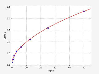
| Human HSP27(Heat Shock Protein 27) ELISA Kit | |
|---|---|
| ELISA Type | Sandwich |
| Sensitivity | 0.469ng/ml |
| Range | 0.781-50ng/ml |
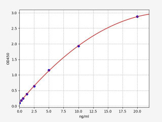
| Human BIM / BCL2L11 ELISA Kit | |
|---|---|
| ELISA Type | Sandwich |
| Sensitivity | 0.188ng/ml |
| Range | 0.313-20ng/ml |
| Human MAPKAPK2 (MAP kinase-activated protein kinase 2) ELISA Kit (HUFI03506) | |
|---|---|
| ELISA Type | Sandwich |
| Sensitivity | 0.094ng/ml |
| Range | 0.156-10ng/ml |
Written by Sean Mac Fhearraigh
Seán Mac Fhearraigh PhD is a co-founder of Assay Genie. Seán carried out his undergraduate degree in Genetics at Trinity College Dublin, followed by a PhD at University College Dublin. He carried out a post-doc at the Department of Genetics, University of Cambridge. Seán is now Chief Technical Officer at Assay Genie.
Recent Posts
-
ELISA vs Western Blot: Which Technique Should You Choose?
Written by Seán Mac Fhearraigh, PhD • Updated: 19 May 2026 • ~9 min read Quick Answer ELISA …20th May 2026 -
Types of ELISA: Direct, Indirect, Sandwich & Competitive Compared
Written by Seán Mac Fhearraigh, PhD • Updated: 19 May 2026 • ~9 min read Quick Answer The fi …20th May 2026 -
Sandwich ELISA: Step-by-Step Protocol & Troubleshooting
Written by Seán Mac Fhearraigh, PhD • Updated: 19 May 2026 • ~ …20th May 2026