
Excluding Doublets in FACS Analysis: A Comprehensive Guide
Excluding Doublets in Flow Cytometry: Pulse Geometry and Gating Strategy
Two cells passing the laser together are recorded as one event, with roughly double the signal. In DNA content analysis a G1 doublet is indistinguishable from a genuine G2/M cell, and in immunophenotyping a doublet can appear double-positive for markers on two different cells. Doublet exclusion rests on one piece of physics — what happens to pulse area, height and width when two cells pass together.
Browse flow cytometry reagents →Key takeaways
- When two similar cells pass the laser together, pulse area roughly doubles and width roughly doubles, while peak height stays close to that of a single cell.
- That asymmetry is the whole basis of doublet discrimination: doublets have high area relative to height, so they fall off the FSC-A versus FSC-H diagonal.
- The opposite statement — that doublets show higher height relative to area — is a common inversion, and gating on it removes the wrong events.
- Standard sequence: FSC-A vs FSC-H first, then FSC-W or SSC-W as a second pass, with fluorescence area versus height where needed.
- Doublets matter most in DNA content work, where a G1 doublet sits exactly where a G2/M single cell should, inflating the apparent G2/M fraction.
- Prevention beats gating: filter immediately before acquisition, avoid over-concentrating, and run at a lower flow rate to reduce coincidence.
- Do not over-gate. Genuinely large single cells — blasts, activated lymphocytes, megakaryocytes — sit near the doublet region and can be discarded by an aggressive gate.
Reagents where doublets distort the result
Doublet exclusion matters most in the assays below — DNA content, apoptosis quadrants and proliferation are all directly corrupted by uncorrected doublets.
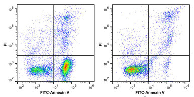
Propidium Iodide Staining Solution
DNA content analysis is the application most damaged by doublets — a G1 pair mimics a G2/M cell exactly.
View product →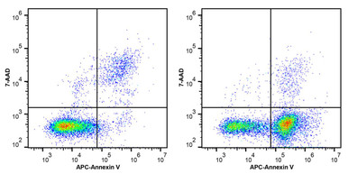
7-AAD Viability Staining Solution
Dead cells and debris promote aggregation, so viability gating and doublet gating work together.
View product →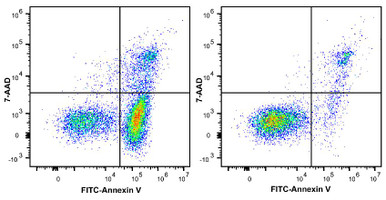
Annexin V-FITC / 7-AAD Apoptosis Kit
Doublets of a live and an apoptotic cell land in the double-positive quadrant and inflate late apoptosis.
View product →
EdU Cell Proliferation Flow Cytometry Assay Kit
S-phase quantification depends on clean singlet gating to avoid counting aggregates as cycling cells.
View product →![FITC Anti-Human CD3 Antibody [OKT-3]](https://cdn11.bigcommerce.com/s-h68l9z2lnx/products/20028/images/606504/fitc-anti-human-cd3-antibody-okt-3-agel0057__97655.1707498205.386.513.jpg?c=2)
FITC Anti-Human CD3 Antibody [OKT-3]
In immunophenotyping, a doublet of two differently stained cells reads as a spurious double-positive.
View product →![FITC Mouse IgG1, Isotype Control [MOPC-21]](https://cdn11.bigcommerce.com/s-h68l9z2lnx/products/229243/images/602526/fitc-mouse-igg1-k-isotype-control-mopc-21-agel2562__43574.1706284986.386.513.jpg?c=2)
FITC Mouse IgG1, Isotype Control [MOPC-21]
A matched isotype control separates non-specific staining from the aggregation artefacts above.
View product →Why doublets matter
A doublet is two cells passing the interrogation point together. The instrument has no way to know it saw two objects; it records one event with the combined signal. The consequences differ by application:
- DNA content analysis is the worst affected. Two G1 cells together carry 2n + 2n = 4n DNA — exactly the content of a genuine G2/M cell. Without doublet exclusion the G2/M fraction is inflated and any cell cycle conclusion is unsafe.
- Immunophenotyping — a doublet of a CD3-positive and a CD19-positive cell reads as a single CD3/CD19 double-positive event, generating a population that does not exist.
- Apoptosis assays — a live cell stuck to an apoptotic one lands in the double-positive quadrant, overstating late apoptosis.
- Rare event detection — when the population of interest is a small fraction of total events, doublet artefacts can rival or exceed the real signal.
- Sorting — a doublet sorted as a target cell delivers an unwanted passenger, which matters for single-cell downstream work.
Pulse geometry: area, height and width
Everything follows from what the detector records as a particle crosses the beam. The signal over time forms a pulse, and the instrument reports three measures of it.
| Parameter | What it measures | Single cell | Doublet |
|---|---|---|---|
| Area (A) | Integrated signal under the whole pulse | Baseline | Roughly double — twice the material passes |
| Height (H) | Peak signal intensity at one instant | Baseline | Approximately unchanged — the beam still sees one cell at a time |
| Width (W) | Duration of the pulse | Baseline | Roughly double — the pair takes twice as long to pass |
The reason height does not double is worth stating explicitly, because it is the crux. Two cells travelling end to end enter the beam one after the other. At any instant the laser illuminates roughly one cell’s worth of material, so the peak is similar to a single cell. What changes is how long the signal lasts — and therefore the total area beneath it.
So a doublet is characterised by a high area-to-height ratio and a wide pulse. Both are directly exploitable.
The common inversion
An earlier version of this guide stated the rationale the other way round — that doublets show a higher peak height relative to area. That is inverted, and it matters because it points the gate at the wrong events.
To be unambiguous: on an FSC-A versus FSC-H plot with area on the x-axis and height on the y-axis, single cells form a tight diagonal because area and height rise together. Doublets have extra area without extra height, so they sit to the right of, and below, the singlet diagonal. The singlet gate is drawn around the diagonal itself; events displaced toward higher area are excluded.
The same logic in reverse also holds: nothing normal produces high height with low area, so that region is generally empty apart from noise.
Step-by-step gating
1. FSC-A versus FSC-H
The primary and usually sufficient gate. Single cells form a linear population along the diagonal; doublets and aggregates deviate toward higher area. Draw the gate around the diagonal population.
2. FSC-W or FSC-H versus FSC-W
A second pass using pulse width. Singlets show a narrow, tight width distribution; doublets show a distinctly broader one. This catches pairs that the area-height plot missed, particularly cells of unequal size.
3. SSC-A versus SSC-H
The same principle applied to side scatter. Useful in heterogeneous samples where granularity differs between populations, and as a cross-check when forward scatter alone leaves ambiguity.
Two passes are conventional. Applying all three is reasonable for cell cycle work or sorting, where a doublet is especially costly.
Fluorescence-based discrimination
The same pulse geometry applies to fluorescence channels, and it is the standard approach for DNA content analysis.
Plotting PI-A against PI-H separates singlets on a diagonal from doublets displaced toward higher area — for exactly the reason above. Two G1 cells give double the integrated fluorescence but a similar peak, so they sit off the line while a true G2/M cell, with genuinely twice the DNA in one nucleus, sits on it.
That distinction is the entire value of the method: area alone cannot separate a G1 doublet from a G2/M singlet, but area against height can.
A worked gating sequence
| Step | Plot | Action |
|---|---|---|
| 1 | FSC-A vs SSC-A | Select the population of interest; exclude debris at low FSC |
| 2 | FSC-A vs FSC-H | Gate the diagonal to retain singlets |
| 3 | SSC-A vs SSC-H | Optional second doublet gate for heterogeneous samples |
| 4 | Viability dye vs SSC-A | Exclude dead cells, which aggregate readily and bind antibody non-specifically |
| 5 | Marker of interest | Analyse, with the gate set using an FMO control |
Order matters. Doublet exclusion belongs early — before viability and marker gating — because an aggregate carrying a dead cell will otherwise contaminate every downstream gate.
Preventing doublets at the bench
Gating removes doublets from the analysis; it does not recover the cells lost to them. Reducing formation in the first place is more efficient.
- Filter immediately before acquisition through a 35–70 µm mesh. This is the single most effective step, and it must be done just before running rather than earlier.
- Do not over-concentrate. Higher cell density raises the probability of coincidence at the interrogation point.
- Run at a lower flow rate. Slower sample delivery narrows the core stream and reduces the chance of two cells arriving together — the standard trade-off against acquisition time.
- Include EDTA in the buffer where the protocol allows. Chelating calcium reduces cadherin-mediated adhesion.
- Handle gently. Over-vigorous pipetting damages cells, and DNA released from lysed cells is highly effective at gluing the rest together.
- Add DNase for samples with substantial cell death, which addresses that free-DNA problem directly.
The risk of over-gating
A tight singlet gate is not automatically better. Large single cells legitimately produce larger pulses, and can sit close to the doublet region — blasts, activated lymphocytes, megakaryocytes and many tumour cells among them.
An aggressive gate drawn on a lymphocyte-dominated sample will quietly discard exactly the population of interest in a leukaemia or activation study. The safeguard is to check what the excluded events are before accepting the gate, rather than tightening until the plot looks neat.
It is also worth recording the singlet percentage. A sudden drop between samples usually signals a preparation problem rather than biology.
Validation and tips
- Check against a known single-cell control — a well-dissociated cell line — to confirm the gate sits where it should.
- Re-optimise per cell type. Gates drawn for lymphocytes will not suit adherent lines or primary tissue digests.
- Use more than one parameter. Area-height with a width plot is more robust than either alone.
- Record the singlet frequency as a quality metric alongside viability.
- Not all instruments report width natively; some derive it from area and height, which can behave differently at the extremes.
Choosing reagents
DNA stains, viability dyes, apoptosis and proliferation kits, and conjugated antibodies with matched isotype controls — the assays where doublet exclusion changes the answer.
Browse flow cytometry reagents →Frequently asked questions
What happens to pulse height in a doublet?
It stays approximately the same as a single cell. Two cells travelling end to end enter the beam one after the other, so the instantaneous peak is similar. Area and width roughly double instead, because the signal lasts twice as long.
Do doublets have higher height relative to area?
No — the reverse. Doublets have higher area relative to height, which is why they sit off the FSC-A versus FSC-H diagonal toward greater area. Gating on the opposite assumption removes the wrong events.
Which plot should I use first?
FSC-A versus FSC-H, gating around the diagonal singlet population. A width-based plot as a second pass catches pairs of unequal size, and SSC equivalents help in heterogeneous samples.
Why do doublets matter so much in cell cycle analysis?
Because two G1 cells together contain 4n DNA — identical to a genuine G2/M cell. Without doublet exclusion the G2/M fraction is inflated, and area-versus-height plotting is the only way to tell them apart.
How do I reduce doublets before acquisition?
Filter through a 35 to 70 micron mesh immediately before running, avoid over-concentrating the sample, use a lower flow rate, include EDTA where the protocol permits, handle gently, and add DNase if there is significant cell death.
Can doublet gating remove cells I want?
Yes. Large single cells such as blasts, activated lymphocytes and megakaryocytes sit near the doublet region, and an over-tight gate discards them. Check what the excluded events actually are before accepting a gate.
Does a lower flow rate really help?
Yes. Slower sample delivery narrows the core stream and reduces the probability of two cells reaching the interrogation point together. The cost is longer acquisition time, which is usually worth paying for rare-event or cell cycle work.
Recent Posts
-
Suspected Plague Death at a Siberian Anti-Plague Institute: What Researchers Should Know
Infectious Disease · News AnalysisSuspected Plague Death at a Siberian Anti-Plague Institute: What …6th Oct 2026 -
LPS ELISA Troubleshooting: The Importance of Sample Nature
Quick answer A researcher measuring circulating lipopolysaccharide (LPS) in human serum …25th Sep 2026 -

Sekundärantikörper wählen: Die Regel, die im Labor wirklich zählt
Sekundärantikörper wählen: Die Regel, die im Labor wirklich zählt Wählen Sie einen Sekundärantikörp …24th Sep 2026