
Innate Immune System Explained: Step-by-Step Process & Key Concepts
Cells of the Innate Immune System: Types, Functions and Markers
The innate immune system responds within minutes, without prior exposure, using cells that recognise conserved microbial patterns rather than specific antigens. This guide covers each major innate cell type — what it does, how it does it, and which surface markers identify it — along with the receptor families that trigger the response and how the cells coordinate with one another and with adaptive immunity.
Browse immune cell markers →Key takeaways
- Innate immunity acts within minutes and needs no prior exposure, because it recognises conserved molecular patterns rather than specific antigens.
- Pattern recognition receptors fall into distinct families — TLRs, NLRs, RLRs and CLRs — each surveying a different cellular compartment.
- Monocytes circulate as precursors and differentiate into macrophages and dendritic cells on entering tissue; tissue-resident macrophages such as microglia and Kupffer cells largely arise separately during development.
- Neutrophils kill by phagocytosis, degranulation, oxidative burst and NET formation — four distinct mechanisms, not one.
- Dendritic cells are the bridge to adaptive immunity, and are the only innate cells that efficiently prime naive T cells.
- NK cells work by missing-self recognition, killing cells that have downregulated MHC class I, and their activity reflects the balance of activating and inhibitory receptors.
- Identifying these populations requires marker combinations — CD68 and CD11b for macrophages, MPO for neutrophils, CD11c with HLA-DR for dendritic cells, CD56 for NK cells.
Contents
- What innate immunity is
- How innate cells recognise threat
- Monocytes
- Macrophages
- Neutrophils
- Eosinophils, basophils and mast cells
- Dendritic cells
- Natural killer cells
- Innate lymphoid and innate-like cells
- Comparison of key innate cells
- Markers for identifying innate cells
- How the cells work together
- References
- Choosing marker antibodies
- Frequently asked questions
Antibodies for innate immune cell identification
No single marker defines an innate cell population. The antibodies below cover one defining marker for each major type, which is the basis of any identification panel.
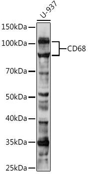
Anti-CD68 Antibody
The classic macrophage marker — a lysosomal glycoprotein widely used to identify tissue macrophages.
View product →![PE Anti-Mouse/Human CD11b Antibody [M1/70]](https://cdn11.bigcommerce.com/s-h68l9z2lnx/products/20186/images/606275/pe-anti-mousehuman-cd11b-antibody-m170-agel0307__86955.1707497481.386.513.jpg?c=2)
PE Anti-Mouse/Human CD11b Antibody [M1/70]
Pan-myeloid integrin marking macrophages, monocytes, neutrophils and granulocytes; cross-reactive across species.
View product →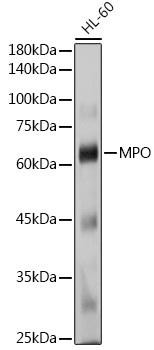
MPO / Myeloperoxidase Antibody
Neutrophil azurophilic granule enzyme — both an identification marker and the enzyme driving the oxidative burst.
View product →![APC Anti-Human CD11c Antibody [BU15]](https://cdn11.bigcommerce.com/s-h68l9z2lnx/products/20320/images/607360/apc-anti-human-cd11c-antibody-bu15-agel0448__84344.1707500940.386.513.jpg?c=2)
APC Anti-Human CD11c Antibody [BU15]
The principal dendritic cell marker, used with HLA-DR to gate conventional DCs.
View product →![FITC Anti-Human CD56 Antibody [5.1H11]](https://cdn11.bigcommerce.com/s-h68l9z2lnx/products/229982/images/602453/fitc-anti-human-cd56-antibody-5.1h11__82570.1706284743.386.513.jpg?c=2)
FITC Anti-Human CD56 Antibody [5.1H11]
NCAM-1, the defining human NK cell marker; CD56-bright and CD56-dim subsets differ functionally.
View product →![PE Anti-Human HLA-DR Antibody [L243]](https://cdn11.bigcommerce.com/s-h68l9z2lnx/products/237258/images/618450/pe-anti-human-hla-dr-antibody-l243-agel0416__17849.1733541274.386.513.jpg?c=2)
PE Anti-Human HLA-DR Antibody [L243]
MHC class II — identifies antigen-presenting cells and pairs with CD11c for the dendritic cell gate.
View product →What innate immunity is
Innate immunity is the arm of the immune system that is present from birth and acts immediately. It does not require prior exposure, does not improve with repetition in the classical sense, and does not generate memory of the kind adaptive immunity produces — although the concept of trained immunity has complicated that last point in recent years.
Its components span physical and chemical barriers — skin, mucosal surfaces, antimicrobial peptides, the complement system — and a set of specialised cells. Those cells are the subject of this guide. The trade-off against adaptive immunity is speed for specificity: innate cells respond within minutes to broad classes of threat, while adaptive responses take days but target a precise antigen.
How innate cells recognise threat
Innate cells carry pattern recognition receptors that detect conserved structures rather than specific epitopes. They respond to two categories of signal: pathogen-associated molecular patterns such as bacterial lipopolysaccharide, flagellin, peptidoglycan and viral nucleic acids; and damage-associated molecular patterns released by injured host cells, including HMGB1, ATP and mitochondrial DNA. The second category is why sterile injury produces inflammation with no infection present.
The receptor families are worth distinguishing, because each surveys a different compartment:
- Toll-like receptors (TLRs) sit on the plasma membrane and in endosomes, detecting bacterial and viral components extracellularly and after uptake.
- NOD-like receptors (NLRs) are cytosolic, sensing intracellular bacterial products and cell stress; several assemble inflammasomes that activate caspase-1 and release IL-1 beta.
- RIG-I-like receptors (RLRs) are cytosolic sensors of viral RNA, driving type I interferon production.
- C-type lectin receptors (CLRs) recognise carbohydrate structures, notably fungal cell wall components.
Engagement of these receptors converges on a small number of signalling pathways — principally NF-kappa-B and the MAPK cascades — producing cytokines, chemokines and interferons.
Monocytes
Monocytes are circulating myeloid cells that act as a mobile reserve. They patrol the bloodstream and, on receiving inflammatory signals, cross the endothelium into tissue where they differentiate into macrophages or dendritic cells. In humans they are divided by CD14 and CD16 expression into classical, intermediate and non-classical subsets, which differ in their patrolling behaviour and inflammatory output.
They matter for interpretation as well as biology: a rise in blood monocytes and a rise in tissue macrophages can reflect the same event, and distinguishing recruited from resident populations usually requires more than one marker.
Macrophages
Macrophages are the principal phagocytes of tissue. They engulf and destroy pathogens, clear dead cells and debris, release cytokines that recruit and instruct other immune cells, and present antigen — though less efficiently than dendritic cells. They also drive tissue repair once infection resolves, which makes them central to both inflammation and its resolution.
Polarisation
Macrophages adopt functional states along a spectrum conventionally summarised as M1 and M2. M1 macrophages, induced by interferon-gamma and LPS, are pro-inflammatory and microbicidal, producing reactive oxygen and nitrogen species and inflammatory cytokines. M2 macrophages, induced by IL-4 and IL-13, favour tissue repair, matrix remodelling and resolution.
The M1/M2 division is a useful shorthand rather than a strict dichotomy — real populations occupy intermediate and mixed states, and the nomenclature has been criticised for implying more discreteness than exists.
Tissue-resident populations
Many tissues host their own long-lived macrophage populations with distinct names and functions: microglia in the central nervous system, Kupffer cells in the liver, alveolar macrophages in the lung, and osteoclasts in bone. A significant point often missed is that these largely arise from embryonic precursors and self-renew in situ, rather than being continuously replaced from blood monocytes.
Neutrophils
Neutrophils are the most abundant circulating leukocyte in humans and the first cells recruited in large numbers to infection. They are short-lived and effectively disposable, arriving in bulk and dying at the site — pus is largely spent neutrophils.
Four killing mechanisms
- Phagocytosis, engulfing opsonised microbes into a phagosome that fuses with granules.
- Degranulation, releasing azurophilic and specific granule contents including myeloperoxidase, elastase, defensins and lactoferrin.
- Oxidative burst, in which NADPH oxidase generates superoxide and myeloperoxidase converts it to hypochlorous acid. Defects in NADPH oxidase cause chronic granulomatous disease.
- NET formation, extruding decondensed chromatin studded with granule proteins to trap microbes extracellularly — a distinct process from phagocytosis, and one implicated in thrombosis and autoimmunity when dysregulated.
Eosinophils, basophils and mast cells
The remaining granulocytes are frequently omitted from summaries of innate immunity, but they carry distinct and non-overlapping roles.
Eosinophils respond to helminth infection and participate in allergic inflammation, releasing major basic protein and eosinophil peroxidase. Basophils are rare in blood and release histamine and IL-4, contributing to allergic responses and to Th2 polarisation. Mast cells are tissue-resident, concentrated at barrier surfaces, and degranulate on IgE cross-linking — the immediate-hypersensitivity reaction. All three are effector cells of type 2 immunity as well as innate defence.
Dendritic cells
Dendritic cells are the bridge between innate and adaptive immunity, and the only innate population that efficiently primes naive T cells. They capture antigen at the periphery, mature on receiving innate signals, migrate to draining lymph nodes, and present processed peptide on MHC alongside costimulatory molecules.
The main subsets are conventional dendritic cells (cDC1 and cDC2), which specialise respectively in cross-presentation to CD8 T cells and in priming CD4 responses; and plasmacytoid dendritic cells, which are weaker antigen presenters but produce large quantities of type I interferon in antiviral responses.
Dendritic cells also maintain tolerance. In the absence of inflammatory signals, presentation of self antigen induces regulatory responses rather than activation — so the same cell type both starts immune responses and prevents inappropriate ones.
Natural killer cells
NK cells are cytotoxic lymphocytes that kill without prior sensitisation and without antigen-specific receptors. They target virally infected and transformed cells, and they contribute to shaping adaptive responses through interferon-gamma production.
Missing-self recognition
The governing principle is a balance of signals. Inhibitory receptors, principally the killer immunoglobulin-like receptors, recognise MHC class I on healthy cells and suppress killing. Activating receptors such as NKG2D and the natural cytotoxicity receptors recognise stress-induced ligands. A cell that has downregulated MHC class I — as viruses and tumours frequently cause — loses inhibitory protection while retaining activating ligands, and is killed. This is missing-self recognition, and it neatly complements cytotoxic T cells, which require MHC class I to act.
Killing proceeds by perforin and granzyme release and by death receptor engagement. NK cells also mediate antibody-dependent cellular cytotoxicity through CD16, which is the mechanism many therapeutic antibodies rely on.
Innate lymphoid and innate-like cells
Beyond NK cells, a broader family of innate lymphoid cells mirrors the helper T cell subsets without antigen-specific receptors: ILC1, ILC2 and ILC3 produce interferon-gamma, type 2 cytokines and IL-17 or IL-22 respectively. They are enriched at barrier surfaces and are important in mucosal defence and tissue homeostasis.
Also innate-like are gamma delta T cells and mucosal-associated invariant T cells, which carry rearranged receptors but recognise a limited, non-peptide set of ligands and respond with innate-like speed. They sit at the boundary between the two arms of immunity and are easily missed in panels designed around conventional T cells.
Comparison of key innate cells
| Cell Type | Main Function | Mechanism of Action | Subtypes |
|---|---|---|---|
| Macrophages | Phagocytosis and cytokine production | Engulfing pathogens; initiating inflammation | M1 (pro-inflammatory), M2 (tissue repair) |
| Neutrophils | Rapid response to infection | Phagocytosis; NET formation | N/A |
| Dendritic Cells | Antigen presentation | Capturing and presenting antigens to T cells | cDCs, pDCs |
| NK Cells | Killing of infected or tumor cells | Inducing apoptosis | N/A |
Markers for identifying innate cells
Identifying these populations experimentally requires marker combinations, since most individual markers are shared across myeloid lineages. The table below gives the commonly used sets.
| Cell type | Human markers | Mouse markers | Notes |
|---|---|---|---|
| Monocytes | CD14, CD16, CD11b, HLA-DR | CD11b, Ly6C, CCR2 | CD14 and CD16 together define the classical, intermediate and non-classical subsets |
| Macrophages | CD68, CD14, CD11b, CD163 (M2) | F4/80, CD11b, MerTK | CD68 is intracellular, so requires permeabilisation for flow cytometry |
| Neutrophils | CD15, CD16, CD66b, MPO | Ly6G, CD11b | Ly6G is the standard mouse neutrophil marker and has no direct human equivalent |
| Dendritic cells | CD11c with HLA-DR; CD123 for pDC | CD11c, MHC II | CD11c alone is insufficient — it is also expressed on macrophages and some other myeloid cells |
| NK cells | CD56, CD16, NKp46, CD3-negative | NK1.1, NKp46, CD3-negative | Excluding CD3 is essential to separate NK cells from NKT and T cells |
| Eosinophils | Siglec-8, CCR3, CD11b | Siglec-F, CCR3 | Siglec-8 and Siglec-F are functional analogues rather than orthologues |
Two practical rules follow. Always include a lineage-exclusion marker — CD3 for NK gating, CD19 for myeloid gating — because contaminating lymphocytes are the commonest source of spurious populations. And where a marker is intracellular, such as CD68 or MPO, fixation and permeabilisation are required, so it cannot be combined naively with a live-cell surface panel.
How the cells work together
The value of the innate system is in the sequence, not in any individual cell.
Tissue-resident macrophages and mast cells detect the initial breach and release cytokines and chemokines. Those signals recruit neutrophils within hours, which arrive in bulk and kill indiscriminately. Monocytes follow, differentiating into macrophages that both continue killing and begin clearing debris. Dendritic cells sample antigen throughout and migrate to lymph nodes, where they prime the adaptive response that arrives days later. NK cells operate in parallel, removing cells that have downregulated MHC to evade cytotoxic T cells.
Resolution is as coordinated as initiation. Macrophages switch toward a repair phenotype, clear apoptotic neutrophils, and release mediators that actively terminate inflammation. Failure of that resolution step — rather than excessive initiation — underlies much chronic inflammatory disease, which is why the M2 transition and apoptotic cell clearance receive as much attention as the killing mechanisms.
References
- Murphy K, Weaver C. Janeway’s Immunobiology. Garland Science — chapters on innate immunity and induced innate responses.
- Iwasaki A, Medzhitov R. Regulation of adaptive immunity by the innate immune system. Science, 2010;327(5963):291–295.
Choosing marker antibodies
Conjugated and unconjugated antibodies against CD68, CD11b, CD11c, CD56, HLA-DR, MPO and the other markers used to identify innate immune populations by flow cytometry and IHC.
Browse immune cell markers →Frequently asked questions
What are the main cells of the innate immune system?
Monocytes, macrophages, neutrophils, eosinophils, basophils, mast cells, dendritic cells and natural killer cells, plus the innate lymphoid cells and innate-like lymphocytes such as gamma delta T cells and MAIT cells.
How does innate immunity differ from adaptive immunity?
Innate immunity acts within minutes using pattern recognition receptors that detect conserved microbial structures, and requires no prior exposure. Adaptive immunity takes days, uses antigen-specific receptors generated by somatic recombination, and produces long-lived memory.
What do pattern recognition receptors detect?
Two things: pathogen-associated molecular patterns such as LPS, flagellin and viral nucleic acids; and damage-associated molecular patterns released by injured host cells, including HMGB1 and mitochondrial DNA. The second category explains why sterile injury causes inflammation.
What is the difference between M1 and M2 macrophages?
M1 macrophages, induced by interferon-gamma and LPS, are pro-inflammatory and microbicidal. M2 macrophages, induced by IL-4 and IL-13, promote tissue repair and resolution. The distinction is a useful shorthand for a spectrum rather than two discrete populations.
How do NK cells know which cells to kill?
By missing-self recognition. Inhibitory receptors detect MHC class I on healthy cells and block killing; activating receptors detect stress ligands. A cell that has downregulated MHC class I loses that protection and is killed — which covers exactly the cells that evade cytotoxic T cells.
Which markers identify macrophages versus neutrophils?
Both express CD11b, so it cannot distinguish them. Macrophages are identified with CD68 or CD163, neutrophils with CD15, CD66b or myeloperoxidase. In mouse, F4/80 marks macrophages and Ly6G marks neutrophils.
Are NETs the same as phagocytosis?
No. Phagocytosis engulfs microbes internally. NET formation extrudes decondensed chromatin coated with granule proteins to trap microbes outside the cell, and the neutrophil typically dies in the process. Dysregulated NET release is implicated in thrombosis and autoimmunity.
Zainab Riaz completed her Master degree in Zoology from Fatimah Jinnah University in Pakistan and is currently pursuing a Doctor of Philosophy in Zoology at University of Lahore in Pakistan.
Recent Posts
-
Suspected Plague Death at a Siberian Anti-Plague Institute: What Researchers Should Know
Infectious Disease · News AnalysisSuspected Plague Death at a Siberian Anti-Plague Institute: What …6th Oct 2026 -
LPS ELISA Troubleshooting: The Importance of Sample Nature
Quick answer A researcher measuring circulating lipopolysaccharide (LPS) in human serum …25th Sep 2026 -

Sekundärantikörper wählen: Die Regel, die im Labor wirklich zählt
Sekundärantikörper wählen: Die Regel, die im Labor wirklich zählt Wählen Sie einen Sekundärantikörp …24th Sep 2026