
Natural Killer Cells: A Guide
Natural Killer Cells: Receptors, Subsets and Function
NK cells kill without prior sensitisation and without antigen-specific receptors, using a balance of activating and inhibitory signals to distinguish healthy cells from infected or transformed ones. This guide covers missing-self recognition, the receptor families that implement it, the CD56-bright and CD56-dim subsets that do quite different jobs, the developmental stages and their markers, and the adaptive NK cells that complicate the textbook claim that NK cells have no memory.
Browse NK cell reagents →Key takeaways
- NK cells are innate lymphocytes that kill without prior sensitisation, guided by the balance between activating and inhibitory receptor signals rather than by antigen specificity.
- Missing-self recognition is the governing principle: inhibitory receptors detect MHC class I on healthy cells, so cells that downregulate it to evade T cells become NK targets instead.
- The two functional subsets differ sharply — CD56-dim CD16-positive cells are the cytotoxic majority in blood, while CD56-bright cells are cytokine producers concentrated in tissue and lymph node.
- CD16 mediates antibody-dependent cellular cytotoxicity, which is how many therapeutic monoclonal antibodies actually kill their targets.
- The claim that NK cells have no immune memory is outdated. Adaptive NK cells, marked by high NKG2C and driven by cytomegalovirus, persist and respond more strongly on re-encounter.
- T cells do not produce antibodies — that is exclusively the function of B cells and plasma cells.
- Development proceeds through defined stages distinguished by CD34, CD117, CD94, CD56 density and CD16, ending in the CD56-dim KIR-positive mature cell.
Contents
- What are natural killer cells?
- NK cells versus T and B cells
- How NK cells kill
- Receptors and missing-self recognition
- CD56-bright and CD56-dim subsets
- Development and stage markers
- Adaptive NK cells and immune memory
- Interactions with other immune cells
- NK cells in health and disease
- Choosing NK reagents
- Frequently asked questions
Reagents for NK cell work
NK identification needs CD56 with CD3 exclusion, and the CD56/CD16 combination to resolve the subsets. The effector readouts below measure what the cells actually did.
![FITC Anti-Human CD56 Antibody [5.1H11]](https://cdn11.bigcommerce.com/s-h68l9z2lnx/products/229982/images/602453/fitc-anti-human-cd56-antibody-5.1h11__82570.1706284743.386.513.jpg?c=2)
FITC Anti-Human CD56 Antibody [5.1H11]
The defining human NK marker; CD56 density separates the bright and dim subsets.
View product →![PerCP/Cyanine5.5 Anti-Human CD16 Antibody [3G8]](https://cdn11.bigcommerce.com/s-h68l9z2lnx/products/229826/images/602870/percpcyanine5.5-anti-human-cd16-antibody-3g8__30145.1706286065.386.513.jpg?c=2)
PerCP/Cyanine5.5 Anti-Human CD16 Antibody [3G8]
Fc gamma receptor IIIa — pairs with CD56 to define the subsets and mediates ADCC.
View product →
Human NKp46 / NCR1 ELISA Kit
A natural cytotoxicity receptor expressed near-exclusively on NK cells across species.
View product →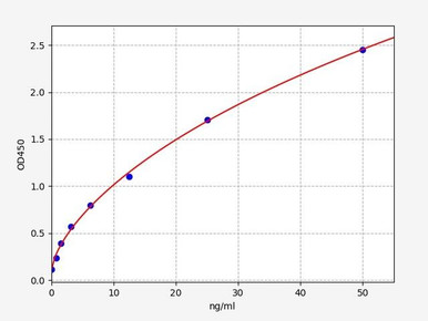
Human Perforin ELISA Kit
The pore-forming protein that admits granzymes into the target cell.
View product →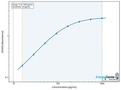
Human Granzyme B ELISA Kit
The principal effector protease of NK killing — a direct measure of degranulation.
View product →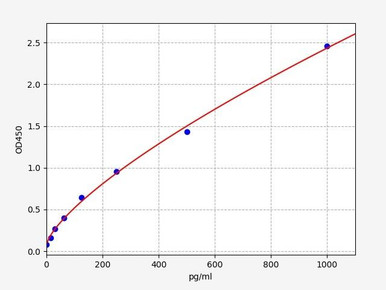
Human IFN-gamma ELISA Kit
The signature CD56-bright output, and the basis of NK cytokine-function assays.
View product →What are natural killer cells?
Natural killer cells are cytotoxic lymphocytes of the innate immune system. They share a common lymphoid progenitor with T and B cells, but unlike those lineages they do not rearrange antigen receptors — there is no NK equivalent of the T cell receptor or antibody. They are called “natural” killers precisely because they need no prior sensitisation: an NK cell encountering a target for the first time can kill it immediately.
Morphologically they are large granular lymphocytes, with abundant cytoplasm containing the cytotoxic granules that store perforin and granzymes. In human blood they make up roughly 5 to 20% of lymphocytes.
NK cells versus T and B cells
| Feature | NK cells | T cells | B cells |
|---|---|---|---|
| Immune arm | Innate | Adaptive | Adaptive |
| Antigen receptor | None rearranged; germline-encoded receptors | T cell receptor, somatically rearranged | B cell receptor / antibody, somatically rearranged |
| Recognition | Balance of activating and inhibitory signals; missing self | Peptide presented on MHC | Native antigen, no MHC required |
| Killing | Direct, via perforin and granzymes, plus ADCC | CD8 cells kill via perforin and granzymes; CD4 cells help | Do not kill; secrete antibody |
| Antibody production | No | No | Yes — as plasma cells |
| Speed | Hours | Days on first exposure | Days on first exposure |
| Memory | Classically none, but adaptive NK cells with memory-like behaviour are now well described | Memory T cells | Memory B cells |
Two rows deserve emphasis because both are commonly stated incorrectly. T cells do not produce antibodies — antibody secretion is exclusive to B cells and their plasma cell progeny, and helper T cells contribute by licensing B cells rather than by making antibody themselves. And the memory row is no longer a clean dichotomy, for reasons covered below.
It is also too simple to divide the labour as NK cells for viruses and cancer, T cells for bacteria. Cytotoxic T cells are central to antiviral immunity, and NK cells contribute to bacterial and parasitic defence largely through interferon gamma.
How NK cells kill
Three mechanisms, used in combination:
- Granule exocytosis. The dominant route. On forming an immunological synapse with a target, the NK cell releases granules containing perforin and granzymes. Perforin polymerises into pores in the target membrane, granzymes enter through them, and granzyme B initiates apoptosis by cleaving caspases and Bid directly.
- Death receptor engagement. NK cells express FasL and TRAIL, which trigger the extrinsic apoptotic pathway in targets expressing the corresponding death receptors.
- Antibody-dependent cellular cytotoxicity. CD16 binds the Fc region of IgG coating a target cell and triggers degranulation. This is not a minor pathway — it is a principal mechanism of action for therapeutic monoclonal antibodies such as rituximab and trastuzumab, which is why CD16 polymorphisms affect clinical response to them.
Alongside killing, NK cells secrete cytokines — principally interferon gamma, which activates macrophages, promotes Th1 differentiation and upregulates MHC class I on surrounding cells. In immunological terms the cytokine output is often as consequential as the cytotoxicity.
Receptors and missing-self recognition
Since NK cells have no antigen-specific receptor, target discrimination rests on integrating signals from two opposing receptor families.
The missing-self principle
Inhibitory receptors recognise MHC class I, which every healthy nucleated cell displays. Engagement delivers a dominant negative signal that prevents killing. Viruses and tumours frequently downregulate MHC class I to escape cytotoxic T cells — and in doing so they remove the NK cell’s inhibitory signal. The two systems are therefore complementary: lose MHC class I and you evade T cells but expose yourself to NK cells.
Inhibition alone is not sufficient, though. A cell must also present activating ligands. Healthy cells that lack MHC class I — erythrocytes, for instance — are not attacked, because there is nothing to trigger activation.
| Receptor family | Type | Ligands recognised |
|---|---|---|
| KIR (CD158 family) | Inhibitory, some activating | HLA-A, -B and -C allotypes; the principal human inhibitory system |
| NKG2A/CD94 | Inhibitory | HLA-E, which displays peptides derived from other HLA molecules — an indirect check on overall class I expression |
| Ly49 family | Inhibitory (mouse) | MHC class I; the functional analogue of human KIRs, though structurally unrelated |
| NKG2D | Activating | MICA, MICB and ULBPs — stress-induced ligands absent from healthy cells |
| Natural cytotoxicity receptors: NKp46 (CD335), NKp30 (CD337), NKp44 (CD336) | Activating | Viral and cellular ligands; NKp44 appears only after activation |
| CD16 (Fc gamma RIIIa) | Activating | IgG Fc bound to a target cell — the ADCC trigger |
The KIR and Ly49 point is worth noting: they perform the same job in humans and mice but are structurally unrelated, an example of convergent evolution. It also means mouse NK data does not map onto human KIR biology directly.
Licensing
NK cells acquire full function through education or licensing: cells whose inhibitory receptors engage self-MHC during development become responsive, while those that cannot engage self-MHC remain hyporesponsive. This is how the system avoids autoreactivity without somatic recombination or thymic selection.
CD56-bright and CD56-dim subsets
Human NK cells divide into two subsets that differ in almost every functional respect, and conflating them is a common source of confusion.
| Feature | CD56-dim | CD56-bright |
|---|---|---|
| Share of blood NK cells | About 90% | About 10% |
| CD16 | High — competent for ADCC | Low or negative |
| Primary function | Cytotoxicity; high perforin and granzyme content | Cytokine production, especially interferon gamma |
| KIR expression | High | Low |
| NKG2A | Variable | High |
| Location | Predominantly blood and spleen | Enriched in lymph nodes, tonsil and other tissue |
| Proliferation | Limited | Responds strongly to IL-2 and IL-15 |
CD56-dim cells are generally regarded as the more mature population, arising from CD56-bright precursors. The practical implication is that measuring total CD56-positive cells conflates a cytotoxic population with a cytokine-producing one — so CD56 and CD16 should be measured together, and CD3 excluded to remove NKT cells.
Development and stage markers
NK cells arise from the common lymphoid progenitor in bone marrow, with lineage commitment governed by transcription factors including Id2, E4BP4 (NFIL3) and T-bet. Development is conventionally divided into stages defined by surface phenotype.
| Stage | Defining phenotype | Character |
|---|---|---|
| Stage 1 (pro-NK) | CD34-positive, CD38-positive, CD117-negative, CD94-negative | Progenitor, not yet NK-committed |
| Stage 2a / 2b (pre-NK) | CD34-positive, CD117-positive; 2a and 2b separated by CD117 and IL-1R1 levels | Committing to the NK lineage |
| Stage 3 (immature NK) | CD34-negative, CD117-positive, CD94-negative; NKG2D, NKp46 (CD335), NKp30 (CD337) and CD161 appearing | Acquiring activating receptors; not yet cytotoxic |
| Stage 4a / 4b | CD94-positive, CD56-bright; NKp80, NKG2D, CD335, CD337 and CD161 at peak | The CD56-bright cytokine-producing population |
| Stage 5 | CD56-dim, CD16 (Fc gamma RIIIa) acquired, KIR (CD158) expression beginning | Mature cytotoxic NK cell |
| Stage 6 | NKG2C-high, expanded and persistent | Adaptive or memory-like NK cells |
The markers that matter are the ones that change between stages — CD34, CD117, CD94, CD56 density, CD16 and KIR. Markers such as CD7 and CD244 are present across most of the sequence and therefore cannot distinguish one stage from the next, which is a common failing of stage tables.
Adaptive NK cells and immune memory
NK cells were long defined partly by what they lacked: no antigen receptor, no clonal expansion, no memory. The first two still hold. The third does not.
Adaptive or memory-like NK cells are now well established. In humans the clearest example is driven by cytomegalovirus infection, which expands a population expressing high levels of the activating receptor NKG2C and lacking the signalling adaptor FcR-gamma. These cells persist for months to years, show distinct epigenetic reprogramming, and respond more vigorously on re-encounter — behaviour that meets any functional definition of memory.
A second form is cytokine-induced memory-like NK cells: brief exposure to IL-12, IL-15 and IL-18 produces cells that respond more strongly weeks later. This is being exploited therapeutically, since such cells can be generated ex vivo before adoptive transfer.
So the flat statement that NK cells have no immune memory should be retired. The accurate position is that they lack antigen-specific memory of the kind mediated by rearranged receptors, while displaying genuine memory-like properties through other mechanisms — part of the broader concept of trained immunity.
Interactions with other immune cells
- Dendritic cells. The interaction is reciprocal: DCs prime NK cells via IL-12, IL-15 and IL-18, while NK-derived interferon gamma promotes DC maturation. NK cells also kill immature DCs, which is thought to serve as quality control on which DCs go on to prime T cells.
- Macrophages. NK-derived interferon gamma is a principal macrophage activator, driving the M1 phenotype and enhancing microbicidal capacity; activated macrophages reciprocate with IL-12 and IL-18.
- T cells. NK cells shape T cell responses through interferon gamma and by editing the DC pool, and they can kill activated T cells to limit responses. Both are cytotoxic lymphocytes, but they recognise targets by entirely different logic.
- B cells and antibody. Through CD16, NK cells are the principal effectors of antibody-dependent cytotoxicity, so the humoral response depends on NK cells to convert antibody binding into target killing.
NK cells in health and disease
Viral infection
NK cells respond early to viral infection, killing infected cells and producing interferon gamma. Their importance is demonstrated by rare NK cell deficiencies, which present with severe and recurrent herpesvirus infections — varicella zoster, cytomegalovirus and Epstein-Barr virus in particular. Many viruses in turn encode proteins that manipulate NK recognition, which is itself evidence of the selective pressure NK cells apply.
Cancer
NK cells contribute to tumour immunosurveillance, targeting cells that downregulate MHC class I or display stress ligands such as MICA and MICB. Tumours evade them by shedding those ligands in soluble form to blunt NKG2D, by secreting TGF-beta and other suppressive factors, and by maintaining sufficient MHC class I to keep inhibitory receptors engaged. Low NK activity has been associated with higher cancer incidence in long-term cohort studies.
Transplantation and pregnancy
In haematopoietic transplantation, KIR-ligand mismatch between donor and recipient can produce a beneficial graft-versus-leukaemia effect, since donor NK cells are not inhibited by recipient MHC. In pregnancy, uterine NK cells are a distinct CD56-bright CD16-negative population that promotes spiral artery remodelling and maternal-fetal tolerance rather than killing — a striking example of the same lineage adapted to an entirely different role.
Autoimmunity
NK involvement in autoimmune disease is genuinely unsettled. They appear to be protective in some contexts, by removing activated immune cells, and pathogenic in others through tissue damage and interferon gamma. Rheumatoid arthritis and multiple sclerosis are both active areas of investigation rather than settled questions.
Choosing NK reagents
CD56 and CD16 antibodies for subset resolution, NKp46 for lineage confirmation, and perforin, granzyme B and interferon gamma assays for measuring cytotoxic and cytokine function.
Browse NK cell reagents →Frequently asked questions
How do NK cells know which cells to kill?
By integrating activating and inhibitory signals. Inhibitory receptors detect MHC class I on healthy cells and block killing; activating receptors detect stress ligands. A cell that has downregulated MHC class I loses that protection — the missing-self principle — and is killed if activating ligands are present.
Do NK cells have immune memory?
Not antigen-specific memory of the T and B cell kind, but the flat claim that they have none is outdated. Adaptive NK cells expressing high NKG2C, expanded by cytomegalovirus, persist and respond more strongly on re-encounter, and cytokine-induced memory-like NK cells behave similarly.
What is the difference between CD56-bright and CD56-dim NK cells?
CD56-dim cells are about 90% of blood NK cells, express CD16 and are the cytotoxic population. CD56-bright cells are around 10%, are CD16-low or negative, and are cytokine producers enriched in lymph node and tissue. Measuring total CD56 conflates the two.
Do T cells produce antibodies?
No. Antibody production is exclusive to B cells and the plasma cells they become. Helper T cells assist B cells in producing antibody but do not make it themselves — a distinction frequently stated incorrectly.
What is ADCC and why does it matter?
Antibody-dependent cellular cytotoxicity: CD16 on the NK cell binds IgG coating a target and triggers killing. It is a principal mechanism of action for therapeutic antibodies such as rituximab and trastuzumab, and CD16 polymorphisms influence how well patients respond to them.
Which markers identify NK cells?
CD56-positive with CD3 excluded is the standard human definition — CD3 exclusion is essential, since CD3-positive CD56-positive cells are NKT cells, not NK cells. CD16 resolves the subsets, and NKp46 is a near-exclusive NK marker across species.
Why do NK cells not kill red blood cells, which lack MHC class I?
Because missing self is necessary but not sufficient. Killing also requires engagement of activating receptors, and erythrocytes present no activating ligands. Both signals must align for cytotoxicity.
Seán Mac Fhearraigh PhD is a co-founder of Assay Genie. Seán carried out his undergraduate degree in Genetics at Trinity College Dublin, followed by a PhD at University College Dublin. He carried out a post-doc at the Department of Genetics, University of Cambridge. Seán is now Chief Technical Officer at Assay Genie.
Recent Posts
-
Suspected Plague Death at a Siberian Anti-Plague Institute: What Researchers Should Know
Infectious Disease · News AnalysisSuspected Plague Death at a Siberian Anti-Plague Institute: What …6th Oct 2026 -
LPS ELISA Troubleshooting: The Importance of Sample Nature
Quick answer A researcher measuring circulating lipopolysaccharide (LPS) in human serum …25th Sep 2026 -

Sekundärantikörper wählen: Die Regel, die im Labor wirklich zählt
Sekundärantikörper wählen: Die Regel, die im Labor wirklich zählt Wählen Sie einen Sekundärantikörp …24th Sep 2026