The MID1IP1 Antibody (PAC056182) is a reliable tool for researchers studying MID1IP1, a protein involved in regulating cell division and proliferation. This polyclonal antibody was raised in rabbits and is highly specific for detecting MID1IP1 in human samples, making it suitable for Western blot applications. By binding to MID1IP1, researchers can analyze its expression levels in different cell types, advancing studies in cell biology and cancer research.MID1IP1, also known as Mid1-interacting protein 1, plays a crucial role in maintaining genomic stability and controlling cell growth. Dysregulation of MID1IP1 has been linked to cancer development and progression, making it a promising target for therapeutic interventions.
By understanding the function of MID1IP1, researchers can uncover new mechanisms underlying cancer biology and potentially develop novel treatment strategies.The MID1IP1 Antibody (PAC056182) offers the reliability and specificity needed for accurate detection of MID1IP1 in research experiments. Its application in Western blotting provides valuable insights into the role of MID1IP1 in cellular processes, offering potential avenues for further exploration in cancer biology and therapeutic development.
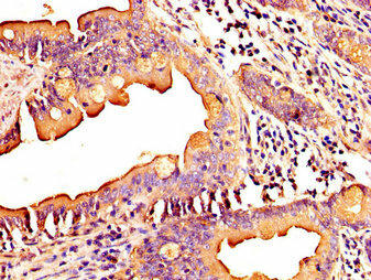
IHC image of PACO56182 diluted at 1:300 and staining in paraffin-embedded human small intestine tissue performed on a Leica BondTM system. After dewaxing and hydration, antigen retrieval was mediated by high pressure in a citrate buffer (pH 6.0). Section was blocked with 10% normal goat serum 30min at RT. Then primary antibody (1% BSA) was incubated at 4°C overnight. The primary is detected by a biotinylated secondary antibody and visualized using an HRP conjugated SP system.
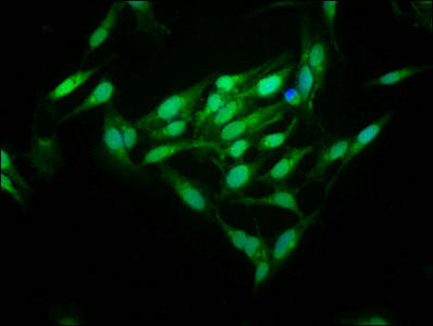
Immunofluorescence staining of Hela cells with PACO56182 at 1:125, counter-stained with DAPI. The cells were fixed in 4% formaldehyde, permeabilized using 0.2% Triton X-100 and blocked in 10% normal Goat Serum. The cells were then incubated with the antibody overnight at 4°C. The secondary antibody was Alexa Fluor 488-congugated AffiniPure Goat Anti-Rabbit IgG(H+L).
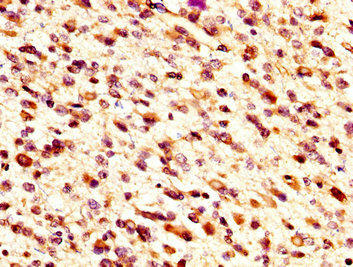
IHC image of PACO56182 diluted at 1:300 and staining in paraffin-embedded human glioma performed on a Leica BondTM system. After dewaxing and hydration, antigen retrieval was mediated by high pressure in a citrate buffer (pH 6.0). Section was blocked with 10% normal goat serum 30min at RT. Then primary antibody (1% BSA) was incubated at 4°C overnight. The primary is detected by a biotinylated secondary antibody and visualized using an HRP conjugated SP system.
Background:
Plays a role in the regulation of lipogenesis in liver. Up-regulates ACACA enzyme activity. Required for efficient lipid biosynthesis, including triacylglycerol, diacylglycerol and phospholipid. Involved in stabilization of microtubules (By similarity).
MID1IP1: Plays a role in the regulation of lipogenesis in liver. Up-regulates ACACA enzyme activity. Required for efficient lipid biosynthesis, including triacylglycerol, diacylglycerol and phospholipid. Involved in stabilization of microtubules. Belongs to the SPOT14 family.Chromosomal Location of Human Ortholog: Xp11.4Cellular Component: cytosol; microtubule cytoskeletonMolecular Function: protein bindingBiological Process: negative regulation of microtubule depolymerization; positive regulation of fatty acid biosynthetic process; positive regulation of ligase activity; protein polymerization; regulation of lipid biosynthetic process


. Section was blocked with 10% normal goat serum 30min at RT. Then primary antibody (1% BSA) was incubated at 4°C overnight. The primary is detected by a Goat anti-rabbit IgG labeled by HRP and visualized using 0.05% DAB.")
.")

.")


. Section was blocked with 10% normal goat serum 30min at RT. Then primary antibody (1% BSA) was incubated at 4°C overnight. The primary is detected by a Goat anti-rabbit polymer IgG labeled by HRP and visualized using 0.05% DAB.")
. Section was blocked with 10% normal goat serum 30min at RT. Then primary antibody (1% BSA) was incubated at 4°C overnight. The primary is detected by a Goat anti-rabbit polymer IgG labeled by HRP and visualized using 0.05% DAB.")