

VEGFR1 (Vascular Endothelial Growth Factor Receptor 1) is a receptor involved in blood vessel formation. The Porcine VEGFR1 ELISA kit allows researchers to gain insights into the role of VEGFR1 in angiogenesis and other processes related to blood vessel formation in pigs, providing valuable information for their studies.
Description
Porcine VEGFR1 / Flt1 / Vascular Endothelial Growth Factor Receptor 1 ELISA Kit
Key Features
| Save Time | Pre-coated 96 well plate | |
| Quick Start | Kit includes all necessary reagents | |
| Publication Ready | Reproducible and reliable results |
Overview
| Product Name: | Porcine VEGFR1/Flt1 (Vascular Endothelial Growth Factor Receptor 1) ELISA Kit |
| Product Code: | PRFI00163 |
| Size: | 96 Assays |
| Alias: | VEGFR1, FLT1, Vascular Endothelial Growth Factor Receptor 1, FLT1, Flt-1, FLT-1, Fms-like tyrosine kinase 1, fms-related tyrosine kinase 1, vascular endothelial growth factor, vascularpermeability factor receptor, FRT, Tyrosine-protein kinase FRT, Tyrosine-protein kinase receptor FLT, vascular endothelial growth factor receptor 1, Vascular permeability factor receptor, VEGFR1, VEGFR-1 |
| Detection Method: | Sandwich ELISA, Double Antibody |
| Reactivity: | Porcine |
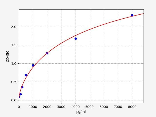
| Sensitivity: | 18.75pg/ml |
| Range: | 31.25-2000pg/ml |
| Storage: | 4°C for 6 months |
| Note: | For Research Use Only |
Additional Information
| Recovery | Matrices listed below were spiked with certain level of Porcine VEGFR1/Flt1R1/Flt1R1/Flt1 and the recovery rates were calculated by comparing the measured value to the expected amount of Porcine VEGFR1/Flt1R1/Flt1R1/Flt1 in samples. Please contact us for more information. |
| Linearity: | The linearity of the kit was assayed by testing samples spiked with appropriate concentration of Porcine VEGFR1/Flt1R1/Flt1R1/Flt1 and their serial dilutions. The results were demonstrated by the percentage of calculated concentration to the expected. Please contact us for more information. |
| CV(%) | Intra Assay <8 Inter Assay <10 |
Kit Components
| Component | Quantity | Storage |
| ELISA Microplate (Dismountable) | 8x12 strips | 2-8°C/-20°C |
| Lyophilized Standard | 2 | 2-8°C/-20°C |
| Sample/Standard Dlution Buffer | 20ml | 2-8°C |
| Biotin-labeled Antibody (Concentrated) | 120ul | 2-8°C (Protection from light) |
| Antibody Dilution Buffer | 10ml | 2-8°C |
| HRP-Streptavidin Conjugate (SABC) | 120ul | 2-8°C(Protect from light) |
| SABC Dilution Buffer | 10ml | 2-8°C |
| TMB Substrate | 10ml | 2-8°C (Protection from light) |
| Stop Solution | 10ml | 2-8°C |
| Wash Buffer (25X) | 30ml | 2-8°C |
| Plate Sealer | 5 | - |
Other materials required:
- Microplate reader with 450 nm wavelength filter
- Multichannel Pipette, Pipette, microcentrifuge tubes and disposable pipette tips
- Incubator
- Deionized or distilled water
- Absorbent paper
- Buffer resevoir
Protocol
*Note: Protocols are specific to each batch/lot. For the exact instructions please follow the protocol included in your kit.
Before adding to wells, equilibrate the SABC working solution and TMB substrate for at least 30 min at 37°C. When diluting samples and reagents, they must be mixed completely and evenly. It is recommended to plot a standard curve for each test.
| Step | Procedure |
| 1. | Set standard, test sample and control (zero) wells on the pre-coated plate respectively, and then, record their positions. It is recommended to measure each standard and sample in duplicate. Wash plate 2 times before adding standard, sample and control (zero) wells! |
| 2. | Aliquot 0.1ml standard solutions into the standard wells. |
| 3. | Add 0.1 ml of Sample / Standard dilution buffer into the control (zero) well. |
| 4. | Add 0.1 ml of properly diluted sample ( Human serum, plasma, tissue homogenates and other biological fluids.) into test sample wells. |
| 5. | Seal the plate with a cover and incubate at 37 °C for 90 min. |
| 6. | Remove the cover and discard the plate content, clap the plate on the absorbent filter papers or other absorbent material. Do NOT let the wells completely dry at any time. Wash plate X2. |
| 7. | Add 0.1 ml of Biotin- detection antibody working solution into the above wells (standard, test sample & zero wells). Add the solution at the bottom of each well without touching the side wall. |
| 8. | Seal the plate with a cover and incubate at 37°C for 60 min. |
| 9. | Remove the cover, and wash plate 3 times with Wash buffer. Let wash buffer rest in wells for 1 min between each wash. |
| 10. | Add 0.1 ml of SABC working solution into each well, cover the plate and incubate at 37°C for 30 min. |
| 11. | Remove the cover and wash plate 5 times with Wash buffer, and each time let the wash buffer stay in the wells for 1-2 min. |
| 12. | Add 90 µl of TMB substrate into each well, cover the plate and incubate at 37°C in dark within 10-20 min. (Note: This incubation time is for reference use only, the optimal time should be determined by end user.) And the shades of blue can be seen in the first 3-4 wells (with most concentrated standard solutions), the other wells show no obvious color. |
| 13. | Add 50 µl of Stop solution into each well and mix thoroughly. The color changes into yellow immediately. |
| 14. | Read the O.D. absorbance at 450 nm in a microplate reader immediately after adding the stop solution. |
Sample Preparation
When carrying out an ELISA assay it is important to prepare your samples in order to achieve the best possible results. Below we have a list of procedures for the preparation of samples for different sample types.
| Sample Type | Protocol |
| Serum | If using serum separator tubes, allow samples to clot for 30 minutes at room temperature. Centrifuge for 10 minutes at 1,000x g. Collect the serum fraction and assay promptly or aliquot and store the samples at -80°C. Avoid multiple freeze-thaw cycles. If serum separator tubes are not being used, allow samples to clot overnight at 2-8°C. Centrifuge for 10 minutes at 1,000x g. Remove serum and assay promptly or aliquot and store the samples at -80°C. Avoid multiple freeze-thaw cycles. |
| Plasma | Collect plasma using EDTA or heparin as an anticoagulant. Centrifuge samples at 4°C for 15 mins at 1000 × g within 30 mins of collection. Collect the plasma fraction and assay promptly or aliquot and store the samples at -80°C. Avoid multiple freeze-thaw cycles. Note: Over haemolysed samples are not suitable for use with this kit. |
| Urine & Cerebrospinal Fluid | Collect the urine (mid-stream) in a sterile container, centrifuge for 20 mins at 2000-3000 rpm. Remove supernatant and assay immediately. If any precipitation is detected, repeat the centrifugation step. A similar protocol can be used for cerebrospinal fluid. |
| Cell culture supernatant | Collect the cell culture media by pipette, followed by centrifugation at 4°C for 20 mins at 1500 rpm. Collect the clear supernatant and assay immediately. |
| Cell lysates | Solubilize cells in lysis buffer and allow to sit on ice for 30 minutes. Centrifuge tubes at 14,000 x g for 5 minutes to remove insoluble material. Aliquot the supernatant into a new tube and discard the remaining whole cell extract. Quantify total protein concentration using a total protein assay. Assay immediately or aliquot and store at ≤ -20 °C. |
| Tissue homogenates | The preparation of tissue homogenates will vary depending upon tissue type. Rinse tissue with 1X PBS to remove excess blood & homogenize in 20ml of 1X PBS (including protease inhibitors) and store overnight at ≤ -20°C. Two freeze-thaw cycles are required to break the cell membranes. To further disrupt the cell membranes you can sonicate the samples. Centrifuge homogenates for 5 mins at 5000xg. Remove the supernatant and assay immediately or aliquot and store at -20°C or -80°C. |
| Tissue lysates | Rinse tissue with PBS, cut into 1-2 mm pieces, and homogenize with a tissue homogenizer in PBS. Add an equal volume of RIPA buffer containing protease inhibitors and lyse tissues at room temperature for 30 minutes with gentle agitation. Centrifuge to remove debris. Quantify total protein concentration using a total protein assay. Assay immediately or aliquot and store at ≤ -20 °C |
| Breast Milk | Collect milk samples and centrifuge at 10,000 x g for 60 min at 4°C. Aliquot the supernatant and assay. For long term use, store samples at -80°C. Minimize freeze/thaw cycles. |
VEGFR1 Background
VEGFR1/ FLT-1 Gene
Vascular endothelial Growth Factor Receptor-1 (VEGFR-1), also known as Fms Related Receptor Tyrosine Kinase 1 (FLT-1), is a protein-coding gene that codes for a member of the Vascular Endothelial Growth Factor receptor (VEGFR) family. Several transcript variants, that encode different isoforms, have been identified for this gene. In Humans, cells that express VEGFR1 include most vascular endothelial cells and peripheral blood monocytes. Tissues where VEGFR1 expression is observed includes lung, placenta, liver, kidney, heart and brain tissues. Specific isoforms can be found in corneal epithelial cells and vascular smooth muscle cells (VSMC).
VEGFR1/ FLT-1 Protein
The VEGFR family of proteins are tyrosine kinase receptors (RTKs). The structure of VEGFR1 contains an extracellular ligand-binding region with seven immunoglobulin (Ig) domains, a transmembrane segment and a cytoplasmic tyrosine kinase (TK) domain. Different isoforms have been identified, including both full-length transmembrane receptor isoforms and shortened, soluble isoforms. The latter forms are associated with the onset of pre-eclampsia
VEGFR-1 Ligands and Function
The VEGFR1 protein acts as a cell surface receptor for VEGF-A, VEGF-B and placental growth factor (PGF). The protein pays an essential role in embryonic vasculature development, angiogenesis regulation, cell migration and survival, chemotaxis and macrophage function and tumour cell invasion. I
Both VEGFR1 and 2 have high binding affinities to VEGF-A. However, VEGFR1 differs from VEGFR2/KDR in its protein kinase activity, which is very low compared to the latter. Thus, VEGFR1 is thought to function as a negative regulator of VEGFA signaling by limiting the amount of free VEGFA and preventing its binding to KDR. The protein also modulates KDR signalling by forming heterodimers.
VEGFR-1 and Angiogenesis
VEGFR1 plays a critical role in angiogenesis. Its interactions with VEGF-A, VEGF-B and PGF initiate signalling pathways such as the MAPK1/ERK2, MAPK3/ERK1 and the MAP kinase signaling pathway, as well as of the AKT1 signaling pathway. However, it is important to note that VEGFR1 exerts both positive and negative regulatory effects on angiogenesis, exhibiting a multifaceted role in vascular development and homeostasis.
While it has been noted to play an essential role as a negative regulator of embryonic angiogenesis by inhibiting excessive proliferation of endothelial cells, they are also noted to promote endothelial cell proliferation, survival and angiogenesis in adulthood.
Its function in promoting cell proliferation seems to be cell-type specific. Promotes PGF-mediated proliferation of endothelial cells, proliferation of some types of cancer cells, but does not promote proliferation of normal fibroblasts (in vitro).The soluble form of VEGFR1, sFlt-1, acts as a decoy receptor that binds and sequesters VEGF ligands,thereby limiting their interaction with membrane-bound VEGFRs.This regulatory mechanism allows VEGFR1 to modulate angiogenesis in a context-dependent manner.
Delving into the intricate mechanisms governing VEGFR1 signaling and its interplay with other VEGF receptors becomes crucial in providing a foundation for developing targeted therapeutic strategies aimed at manipulating angiogenesis in various pathological conditions.
VEGFR-1 Inhibitors
VEGFR inhibitors are a class of cancer drugs that can inhibit angiogenesis by inhibiting the VEGF pathway. Both small molecules as well as monoclonal antibodies have been developed as VEGFR-1 inhibitors. VEGFR1 inhibitors may exhibit varying degrees of selectivity and may also affect other receptors, such as VEGFR2 and VEGFR3. Some examples are Axitinib (Inlyta), Pazopanib (Votrient) and Cediranib (Recentin).
Porcine VEGFR1/FLT1 ELISA Kit FAQs
Q: What is the purpose of a Porcine VEGFR1 ELISA Kit?
The Porcine VEGFR1/FLT1 ELISA Kits are designed to measure the levels of Vascular Endothelial Growth Factor Receptor 1 (VEGFR1) in porcine samples. This allows researchers to investigate the role of VEGFR1 in angiogenesis and other processes related to blood vessel formation in pigs.
Q: What type of samples can be used with a Porcine VEGFR1 ELISA Kit?
Porcine VEGFR1 ELISA Kits are compatible with a wide range of sample types, including porcine serum, plasma, cell lysates, and tissue homogenates. Researchers can use these kits to measure VEGFR1 levels in various porcine biological fluids and tissues.
Q: Are Porcine VEGFR1 ELISA Kits specific to porcine samples only?
Yes, Porcine VEGFR1 ELISA Kits are specifically designed and validated for the detection and quantification of VEGFR1 in porcine samples. They are not recommended for use with samples from other species unless cross-reactivity has been explicitly confirmed.
Q: Where can I find additional technical support or assistance with the Porcine VEGFR1 ELISA kit?
For any technical inquiries or assistance regarding the Porcine VEGFR1 ELISA kit, you can reach out to our team. They will be available to answer your questions and provide the necessary guidance to ensure a successful experiment.
Related Products
| Human VEGFR1 / Flt-1 ELISA Kit | |
|---|---|
| ELISA TYPE: | Sandwich ELISA, Double Antibody |
| SENSITIVITY: | 75pg/ml |
| RANGE: | 125-8000pg/ml |
 ELISA Kit (MOES01155)")
 ELISA Kit")


")