The TTLL3 Polyclonal Antibody (PACO59960) is a valuable tool for researchers studying TTLL3, a protein involved in the post-translational modification of tubulins. This antibody, produced in rabbits, is highly specific to human TTLL3 and has been validated for use in Western blot applications. By binding to the TTLL3 protein, this antibody allows for the detection and analysis of TTLL3 expression in various cell types, making it an ideal choice for studies in the fields of molecular biology and cell biology.TTLL3 is known to play a crucial role in the modification of tubulins, which are essential components of the cytoskeleton and play a key role in cell division and intracellular transport.
Dysregulation of tubulin modifications has been linked to various diseases, including cancer and neurological disorders. Therefore, understanding the function of TTLL3 is important for unraveling the molecular mechanisms underlying these conditions and developing targeted therapies.By using the TTLL3 Polyclonal Antibody, researchers can explore the role of TTLL3 in cellular processes and disease pathways, furthering our understanding of tubulin modification and its implications for human health. This antibody is a valuable tool for studies aimed at unraveling the intricate molecular mechanisms governing cell biology and disease progression.
Antibody Name:
TTLL3 Antibody (PACO59960)
Antibody SKU:
PACO59960
Size:
50ug
Host Species:
Rabbit
Tested Applications:
ELISA, IHC
Recommended Dilutions:
ELISA:1:2000-1:10000, IHC:1:200-1:500
Species Reactivity:
Human
Immunogen:
Recombinant Human Tubulin monoglycylase TTLL3 protein (470-641AA)
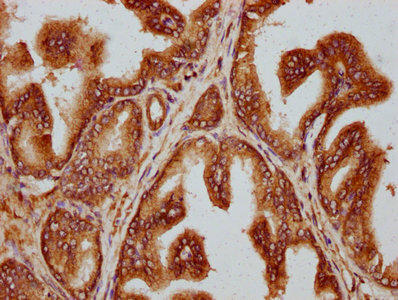
IHC image of PACO59960 diluted at 1:300 and staining in paraffin-embedded human prostate cancer performed on a Leica BondTM system. After dewaxing and hydration, antigen retrieval was mediated by high pressure in a citrate buffer (pH 6.0). Section was blocked with 10% normal goat serum 30min at RT. Then primary antibody (1% BSA) was incubated at 4°C overnight. The primary is detected by a biotinylated secondary antibody and visualized using an HRP conjugated SP system.
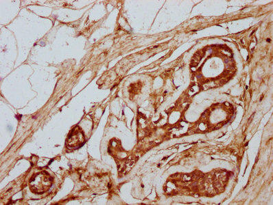
IHC image of PACO59960 diluted at 1:300 and staining in paraffin-embedded human breast cancer performed on a Leica BondTM system. After dewaxing and hydration, antigen retrieval was mediated by high pressure in a citrate buffer (pH 6.0). Section was blocked with 10% normal goat serum 30min at RT. Then primary antibody (1% BSA) was incubated at 4°C overnight. The primary is detected by a biotinylated secondary antibody and visualized using an HRP conjugated SP system.
Background:
Monoglycylase which modifies alpha- and beta-tubulin, generating side chains of glycine on the gamma-carboxyl groups of specific glutamate residues within the C-terminal tail of alpha- and beta-tubulin. Involved in the side-chain initiation step of the glycylation reaction by adding a single glycine chain to generate monoglycine side chains. Not involved in elongation step of the polyglycylation reaction.


.")
.")
. Section was blocked with 10% normal goat serum 30min at RT. Then primary antibody (1% BSA) was incubated at 4°C overnight. The primary is detected by a Goat anti-rabbit IgG labeled by HRP and visualized using 0.05% DAB.")
.")
.")

.")
.")