
Blog
Treg Suppression and the IL-2 Axis: Pathway, Function and Assays
Regulatory T cells cannot make the cytokine they depend on. FOXP3 represses the Il2 locus, so every Treg lives on IL-2 secreted by the conventional T cells it exists to restrain. That asymmetry explains most of what follows: why Tregs carry the highest-affinity IL-2 receptor in the body, why STAT5 sits at the centre of the map, and why the axis fails in two directions — autoimmunity when it is too weak, tumour tolerance when too strong.
Key takeaways
Tregs are obligate IL-2 consumers, not producers: FOXP3 with NFAT shuts down Il2 while switching on Il2ra and Ctla4.
CD25 has no signalling tail; it raises the affinity of the IL-2Rβ–γc pair about 100-fold — the bas
…
19th Aug 2026
In Vivo Cell Depletion: Antibody Targets, Fc Mechanisms and Dosing
A depletion experiment is only as good as the depletion you can prove. Injecting a functional-grade antibody and assuming the population is gone is the most common failure mode in in vivo immunology. The marker sets which cells are targeted, the Fc region decides whether they are lysed, eaten or merely coated, and the tissue decides how complete the loss really is. This atlas maps ten validated depleting targets onto the populations they remove and onto the three effector mechanisms that do the work.
Key takeaways
Marker choice sets specificity: Ly6G is neutrophil-selective, while Gr-1 (Ly6G plus Ly6C) also removes monocytes and MDSC.
Three mechanisms clear the opsonised cell: ADCC (NK Fc&g
…
19th Aug 2026
T-Cell Co-Stimulation and Agonist Checkpoint Signalling Explained
Signal 1 tells a T cell what it is looking at; signal 2 tells it whether to care. The same peptide–MHC engagement that launches a productive effector response can just as easily install an anergy programme, and the difference is decided by a second set of receptors acting over the following hours and days. Two structurally unrelated systems do that work — the CD28 family and the TNF-receptor superfamily — and they are not redundant. Reagents that engage them behave nothing like the blocking antibodies most of us are used to.
Key takeaways
NFAT without AP-1 is not a weak signal but a different one, transcribing an active anergy programme instead of the IL-2 response.
CD28 w
…
19th Aug 2026
PD-1/PD-L1 Checkpoint Signalling: Pathway, Function and Assays
PD-1 does not switch a T cell off; it moves the threshold. Engagement of PD-1 by PD-L1 recruits a single phosphatase into the immunological synapse, and that one recruitment event is enough to collapse calcium flux, ERK activation and the metabolic programme that sustains an effector response. Checkpoint blockade has made the axis famous, but the mechanism is more selective than most summaries admit — and knowing which substrate is dephosphorylated changes how you design a blocking experiment.
Key takeaways
PD-1 signals through an ITSM, not the adjacent ITIM; phospho-ITSM binds both SH2 domains of SHP-2 and relieves its autoinhibition.
The dominant substrate is CD28, not the TCR &mdas
…
19th Aug 2026
TIGIT, DNAM-1 and the CD155 Axis: Pathway, Function and Assays
CD155 is read by four different receptors, and only one of them turns the killing programme on. The nectin and nectin-like ligands induced on stressed and transformed cells are simultaneously an activating cue through DNAM-1 (CD226) and an inhibitory cue through TIGIT, CD96 and PVRIG. Which signal wins is decided by receptor affinity, by surface stoichiometry, and by a cis-interaction on the effector membrane that has nothing to do with the ligand at all. That balance sets whether an NK or CD8⁺ T cell degranulates.
Key takeaways
CD155 (PVR) and CD112 (nectin-2) are stress-induced ligands read by one activating receptor, DNAM-1/CD226, and three brakes: TIGIT, CD96 and PVRIG.
TIGIT bind
…
19th Aug 2026
NK Cell Cytotoxicity Pathway: Receptors, Signalling and Assays
A natural killer cell decides by subtraction. It carries no rearranged antigen receptor and no memory of what it has seen. It presses a panel of germline-encoded receptors against a target and integrates the result: NKG2D, NKp46, DNAM-1 and CD16 pushing one way, inhibitory KIR and NKG2A pushing the other. If the sum crosses threshold, the cell polarises its granules and delivers perforin and granzyme B within minutes. Everything interesting about NK biology sits in how that sum is computed.
Key takeaways
NK cells kill by arithmetic, not recognition — the balance of activating and inhibitory receptors at one contact sets the outcome.
Missing-self surveillance runs through KIR (human) a
…
19th Aug 2026
NLRP3 Inflammasome and Pyroptosis: Pathway, Function and Assays
The inflammasome is one of the few innate pathways that destroys the cell it activates. A macrophage that assembles NLRP3 has committed to a decision it cannot reverse: a priming signal through TLR4 and NF-κB stocks the cytosol with inactive precursors, then an entirely separate second signal nucleates an ASC speck, activates caspase-1 and punches gasdermin-D pores through its own membrane. The output — IL-1β and IL-18 leaving through those pores — drives fever, neutrophil recruitment and, when the brakes fail, hereditary autoinflammatory disease.
Key takeaways
NLRP3 needs two signals: transcriptional priming via TLR4 → NF-κB, then a separate activation tri
…
19th Aug 2026
B-Cell Receptor Signalling and the Germinal Centre Reaction
The germinal centre is where antibody quality is made. A naive B cell that binds antigen does not become a source of protective, high-affinity IgG by accident. It passes through a tightly choreographed sequence: receptor signalling calibrated by three separate brakes, survival signals from the TNF superfamily, a licensing handshake with a T-follicular-helper cell, and then weeks of iterative mutation and selection inside a specialised anatomical structure. Understanding that sequence is the difference between a vaccine adjuvant that works and one that does not — and between an autoimmune model that phenocopies human disease and one that does not.
Key takeaways
The B-cell receptor has
…
19th Aug 2026
ELISA Troubleshooting: A Low Standard Curve and Samples Reading Off-Scale
Quick answer
On the Periaxin (PRX) ELISA, a researcher found every serum sample reading above the top standard — optical density (OD) around 2.8–3.0 at 450 nm, well beyond the highest calibrator. The kit was not faulty. The samples simply contained far more analyte than a 1:6 dilution could bring into the assay’s 0.156–10 ng/mL range. The fix is to confirm the standard curve, then dilute samples enough (here, closer to 1:100–1:1000) to land them inside the linear region.
On this page
Click any section below to jump straight to it ↓
The problem ›
Snapshot of the data ›
Why it ha
…
12th Aug 2026
FAO Activity Negative Results? Data Analysis Help
Quick answer
On the Fatty Acid Oxidation (FAO) Assay Kit (BR00001), a researcher found that a subset of skeletal-muscle samples returned negative values once the control well was subtracted from the reaction well, while other samples on the same plate read clearly positive. The negatives were not a kit failure: they mark samples whose FAO activity sits at or below the assay's detection floor, where ordinary well-to-well optical noise tips the background-subtracted value just below zero. The fix is interpretation, not a repeat run — report these as ND (not detectable), confirm blood was washed from the tissue, and let the valid positive samples validate the plate.
…
11th Aug 2026
Questions About Dilutions? A Real Example of How to Prepare Your ELISA Solutions
Quick answer
A researcher preparing a Human IL-18 ELISA wasn't sure whether the protocol's “300-fold” HRP-Streptavidin step meant adding 40 µL of concentrate to 12 mL of diluent, or to a total of 12 mL. A fold dilution always describes the final total volume, not the diluent alone. For a 300-fold dilution you combine 1 part concentrate (40 µL) with 299 parts diluent (11,960 µL) to reach 12 mL total — so 40 µL HRP-Streptavidin + 11,960 µL Assay Diluent = 12 mL. The worked example's rounded “12 mL diluent” wording was the source of the confus
…
6th Aug 2026
Red Blood Cell Peroxidase Interference in Competitive ELISA
QUICK ANSWER
A researcher ran a competitive 2,3-bisphosphoglycerate (2,3-BPG) ELISA on red blood cell (RBC) lysate. The standard curve was textbook, yet every lysate sample read at the top of the curve with no dose-response, and the most concentrated sample gave a negative absorbance. The kit was working — the sample matrix was not. RBC lysate carries endogenous peroxidase activity that reacts with the kit’s TMB substrate independently of the HRP conjugate, generating non-specific colour and unreliable readings. RBC lysate is not a validated matrix for this kit, so it needs validation before the numbers can be trusted.
On this page
The pro
…
4th Aug 2026
Sample Dilution Range Calculation for Competitive ELISA kits
Quick answer
A competitive ELISA is running correctly when the blank (zero standard) gives the highest OD, the standard curve drops smoothly with rising concentration, and your samples land in the middle of that curve. If your interpolated values already sit comfortably inside the kit's range — for the Bovine PGE2 (PGE2) ELISA that is 15.6–1000 pg/mL — you generally do not need to dilute. Fit the standards with a 4-parameter logistic (4PL) regression and keep duplicate CVs at or below 20%.
On this page
The problem
The data
Why it happens
Our analysis
Root cause
What we recommended
Key takeaway
FAQ
The problem
A researcher reached out before starting a new
…
31st Jul 2026
Troubleshooting high ELISA concentrations
QUICK ANSWER
If your ELISA returns sample concentrations that are orders of magnitude too high, the assay itself is usually fine — the error is almost always in data analysis. The most common culprit is entering the wrong standard concentrations when building the standard curve. In a recent porcine MAP (PigMAP) sandwich ELISA case, a researcher assigned a 5000–78 ng/mL range to standards that should have been 10–0.156 ng/mL. The optical density (OD) readings were valid; only the concentrations tied to them were wrong. Re-assigning the correct values to the same ODs brought results back into a sensible range.
On this page
The problem
Snapshot of t
…
30th Jul 2026
ELISA Troubleshooting: When Standards Read Below the Blank
Quick answer
On the GDF-6 (Growth Differentiation Factor 6) ELISA Kit, a researcher saw an elevated blank (OD ≈ 0.41) with the two lowest standards reading at or below it, and no signal for a recombinant GDF-6 sample. The cause was a background/handling issue — most likely an over-concentrated HRP conjugate, incomplete washing, or stale wash buffer — not a fault in the antigen or kit. Fresh reagents, thorough washing, and a rebuilt low-end curve resolve it.
On this page
Click any section below to jump straight to it ↓
The problem ›
Snapshot of the data ›
Why it happens ›
Our a
…
28th Jul 2026
Introducing BioSymposia: The Gathering Place for Science
Announcement · Powered by Assay GenieIntroducing BioSymposia: The Gathering Place for ScienceWe’re excited to announce the launch of BioSymposia, our new sister platform and a free home for scientific events. BioSymposia helps researchers discover conferences, symposia and workshops across every discipline — and helps organisers put their events in front of a global research community.Explore BioSymposia →96+events listed25+scientific disciplines80+countries coveredFreeto list your eventKey TakeawaysBioSymposia is Assay Genie’s new sister site: a free platform for discovering scientific events worldwide.Browse conferences, symposia and workshops across 25+ disciplines in 80+ coun
…
21st Jul 2026
Types of ELISA: Direct, Indirect, Sandwich & Competitive Compared
Written by Seán Mac Fhearraigh, PhD • Updated: 19 May 2026 • ~9 min read
Quick Answer
The five main types of ELISA are sandwich, competitive, direct, indirect and multiplex. Sandwich ELISA is the most common — used for proteins and cytokines. Competitive ELISA suits small molecules like hormones. Indirect ELISA is the standard for antibody screening. Direct ELISA is the simplest format. Multiplex measures many analytes per well.
On this page
All ELISA types at a glance
Sandwich ELISA
Competitive ELISA
Direct ELISA
Indirect ELISA
Multiplex ELISA
Other formats (ELISpot, FluoroSpot, CLIA, cell-based)
How to choose the right format
Kit formats: pre-coated vs DIY
…
20th May 2026
Sandwich ELISA: Step-by-Step Protocol & Troubleshooting
Written by Seán Mac Fhearraigh, PhD •
Updated: 19 May 2026 •
~10 min read
Quick Answer
A sandwich ELISA uses two antibodies — a capture antibody immobilised on a 96-well plate and an enzyme-linked detection antibody — to bind two different epitopes on the same target protein. Signal is directly proportional to analyte concentration. A typical sandwich ELISA takes 3–4 hours and detects analyte at pg/mL to ng/mL levels.
On this page
What is a sandwich ELISA?
When to use a sandwich ELISA
The principle in detail
Pre-coated sandwich ELISA protocol
DIY sandwich ELI
…
20th May 2026
ELISA vs Western Blot: Which Technique Should You Choose?
Written by Seán Mac Fhearraigh, PhD • Updated: 19 May 2026 • ~9 min read
Quick Answer
ELISA and Western Blot are both antibody-based protein detection methods. ELISA is faster, higher throughput and gives you a precise quantitative number from complex samples. Western Blot separates proteins by size first, so it tells you whether your antibody is binding the right molecular weight band — making it the gold standard for protein identity confirmation. Use ELISA to measure how much; use Western Blot to confirm what.
On this page
ELISA vs Western Blot at a glance
What is ELISA?
What is Western Blot?
Key differences in detail
Sensitivity & quantitation
Throug
…
20th May 2026
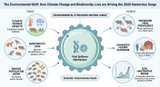
The Environmental Shift: How Climate Change and Biodiversity Loss are Driving the Hantavirus Surge
The year 2026 has brought an unsettling spotlight onto an old adversary: Hantavirus. What began as isolated cases has rapidly escalated into a multi-country surge, leaving scientists and public health officials scrambling for answers. This isn't merely a resurgence; it's a stark warning, revealing how our planet's changing climate and dwindling biodiversity are directly fueling new patterns of disease emergence.
Introduction
Hantaviruses, a family of RNA viruses primarily transmitted to humans through contact with infected rodent excreta, have long posed a public health threat. However, the current global surge in Hantavirus infections is unprecedented, prompting urgent investigations into
…
12th May 2026
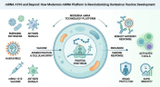
mRNA-1010 and Beyond: How Moderna's mRNA Platform is Revolutionizing Hantavirus Vaccine Development
Hantavirus, a silent threat lurking in rodent populations, has long posed a significant public health challenge with its severe and often fatal outcomes. For decades, the quest for effective vaccines has been a scientific priority. Now, a new era of vaccine development is dawning, spearheaded by revolutionary mRNA technology, promising unprecedented speed and efficacy in our fight against this elusive pathogen.
Introduction
Orthohantaviruses, commonly known as hantaviruses, are a group of zoonotic viruses responsible for two severe human diseases: Hemorrhagic Fever with Renal Syndrome (HFRS) in Eurasia and Hantavirus Pulmonary Syndrome (HPS) in the Americas. These illnesses carry high fata
…
12th May 2026

Prevention and Risk Mitigation: Strategies to Reduce Hantavirus Exposure in a Changing World
As hantavirus cases continue to rise globally, the question becomes not just how to detect infections, but how to prevent them in the first place. Recent epidemiological investigations across California, Qingdao, and the China-Russia border reveal that hantavirus prevention is achievable through a combination of environmental management, public awareness, and targeted surveillance. This comprehensive approach—integrating rodent control, habitat modification, and early detection—offers hope in mitigating the burden of this deadly virus in our changing world.
Introduction
Hantavirus infections have plagued human populations for decades, yet remarkably, the majority of cases are preventable.
…
12th May 2026
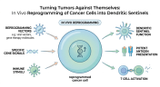
Turning Tumors Against Themselves: In Vivo Reprogramming of Cancer Cells into Dendritic Sentinels
Imagine a future where cancer cells, once masters of disguise, are forced to reveal themselves and even train the body's immune system to fight back. This isn't science fiction; it's the groundbreaking reality emerging from the latest advancements in cancer immunotherapy. Researchers are now exploring a revolutionary strategy: turning tumors against themselves by reprogramming their very cells into powerful immune sentinels.
Introduction
For decades, the fight against cancer has been a relentless pursuit, marked by both triumphs and formidable challenges. While traditional treatments like chemotherapy and radiation have saved countless lives, they often come with significant side effects an
…
3rd Apr 2026
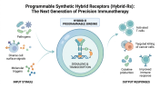
Programmable Synthetic Hybrid Receptors (Hybrid-Rs): The Next Generation of Precision Immunotherapy
Imagine a world where cancer treatment is no longer a blunt instrument but a precision-guided surgical strike, executed by the body's own immune system. For years, we have dreamed of 'smart' cells that can navigate the complex terrain of a tumor, distinguish friend from foe with absolute certainty, and adapt their behavior in real-time. Today, that dream is rapidly becoming a reality through the emergence of Programmable Synthetic Hybrid Receptors, or Hybrid-Rs, which are redefining the very boundaries of immunotherapy.
Introduction
The landscape of modern oncology has been fundamentally altered by the advent of Chimeric Antigen Receptor (CAR) T-cell therapy. By genetically engineering a pa
…
31st Mar 2026