
A Quick Guide to Myelin
A Quick Guide to Myelin
Myelin is the lipid-rich insulating sheath wrapped around nerve axons. Made by oligodendrocytes in the central nervous system and Schwann cells in the periphery, it lets electrical impulses jump between Nodes of Ranvier — saltatory conduction, which makes nerve signalling up to 100 times faster. When myelin is lost, as in multiple sclerosis, conduction slows or fails altogether.
Explore myelin marker ELISA kits →Key Takeaways
- Myelin is a white, fatty membrane of lipids and proteins that insulates axons.
- Oligodendrocytes myelinate the CNS; Schwann cells myelinate the PNS.
- Nodes of Ranvier enable saltatory conduction, dramatically speeding nerve impulses.
- Core myelin proteins — MBP, PLP1, MOG, MAG and CNPase — are the biomarkers used to track myelin integrity.
- Demyelination underlies multiple sclerosis, Guillain-Barré syndrome and the leukodystrophies.
- Remyelination is possible, but becomes inefficient with age and chronic inflammation.
Contents
- What is Myelin?
- Who Makes Myelin? Oligodendrocytes vs Schwann Cells
- Neuron Anatomy and the Role of Axons
- Nodes of Ranvier and Saltatory Conduction
- The Key Myelin Proteins
- Myelin Damage: Causes and Consequences
- Demyelination and Multiple Sclerosis
- Remyelination and Myelin Restoration
- Frequently Asked Questions
Myelin Marker ELISA Kits
These validated sandwich ELISA kits quantify the structural myelin proteins and axonal-damage markers used across demyelination and remyelination research.
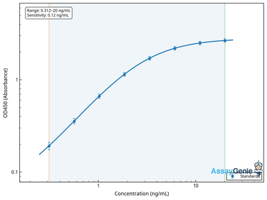
Human MBP ELISA Kit
Myelin basic protein compacts the sheath and is the classic marker of active myelin breakdown in CSF and serum.
View kit →
Human PLP1 ELISA Kit
Proteolipid protein 1 is the single most abundant myelin protein, holding adjacent membrane wraps together.
View kit →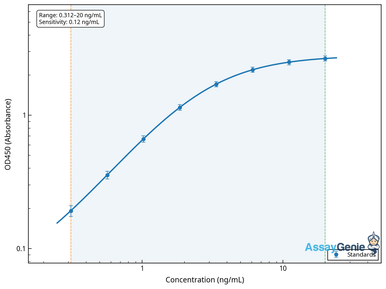
Human MOG ELISA Kit
Myelin oligodendrocyte glycoprotein sits on the outermost sheath surface and is the target antigen in MOGAD.
View kit →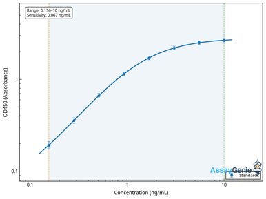
Human MAG ELISA Kit
Myelin-associated glycoprotein mediates the axon–glia contact that maintains the sheath.
View kit →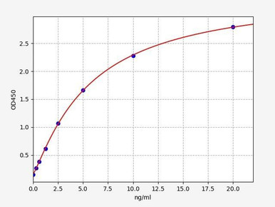
Human CNPase ELISA Kit
CNPase is an oligodendrocyte-enriched enzyme widely used as a marker of myelinating capacity.
View kit →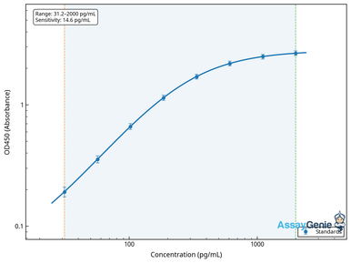
Human NEFL ELISA Kit
Neurofilament light chain reports the axonal damage that follows sustained demyelination.
View kit →What is Myelin?
Myelin is a specialised, tightly compacted extension of the glial cell membrane that spirals around the axon of a nerve cell to form an insulating sheath. Unlike most biological membranes, myelin is dominated by lipid: roughly 70 to 85 per cent of its dry weight is lipid, chiefly cholesterol, galactocerebroside and sphingomyelin, with the remaining fraction made up of a small number of highly abundant proteins. That unusual composition gives myelin both high electrical resistance and low capacitance, and it is also what makes it appear white — which is why myelin-rich regions of the brain and spinal cord are called white matter, in contrast to the cell-body-rich grey matter.
Functionally the sheath does three jobs at once. It insulates the axon so current is not lost across the membrane, it prevents electrical crosstalk between neighbouring fibres, and it provides metabolic support to the axon it wraps — oligodendrocytes shuttle lactate and pyruvate into the axon through monocarboxylate transporters, sustaining fibres far too long to be supplied by the neuronal cell body alone. Myelin is therefore not passive packaging; it is an active partner in neuronal survival, which is why demyelination is so often followed by axonal degeneration.
Who Makes Myelin? Oligodendrocytes vs Schwann Cells
Two entirely different glial cell types build myelin, one in each half of the nervous system. In the brain and spinal cord the job belongs to oligodendrocytes, which extend multiple processes and can myelinate up to 50 separate axon segments at once. In the peripheral nervous system Schwann cells do the work, but each wraps a single internode on a single axon. That difference matters clinically: one dying oligodendrocyte strips myelin from dozens of axons simultaneously, which is part of why CNS demyelinating disease is so disabling.
| Feature | Oligodendrocyte | Schwann cell |
|---|---|---|
| Location | Central nervous system | Peripheral nervous system |
| Axons myelinated per cell | Up to ~50 internodes | One internode |
| Dominant proteins | PLP1, MBP, MOG, MAG, CNPase | P0 (MPZ), PMP22, MBP |
| Regenerative capacity | Limited; declines with age | Robust; supports nerve regrowth |
| Representative disease | Multiple sclerosis | Guillain-Barré syndrome, CMT |
Neuron Anatomy and the Role of Axons
The axon is the long, cable-like projection that carries the action potential away from the neuron cell body toward the axon terminal, where neurotransmitter is released onto the next cell. Dendrites collect incoming signals at one end of the neuron, the cell body integrates them, and the axon transmits the result — sometimes over a metre, as in the motor neurons running from the spinal cord to the foot. Myelin wraps the axon in discrete segments called internodes.
Axon diameter, internode length and sheath thickness are tuned together, and all three influence conduction velocity. The ratio of axon diameter to total fibre diameter — the g-ratio — sits near 0.6 to 0.7 in healthy CNS white matter, and a rising g-ratio is a reliable sign that myelin is thinning.
Nodes of Ranvier and Saltatory Conduction
Myelin does not run continuously along the axon. Every millimetre or so it is interrupted by a short, bare gap of about one micrometre known as a Node of Ranvier. These nodes are densely packed with voltage-gated sodium channels, while the myelinated internodes between them have very few. The action potential therefore cannot regenerate under the sheath — current flows passively down the insulated internode and the impulse is regenerated only at the next node, appearing to leap from node to node. This is saltatory conduction, from the Latin saltare, to jump.
The payoff is enormous. An unmyelinated axon conducts at roughly 0.5 to 2 metres per second; a myelinated axon of the same diameter conducts at up to 120 metres per second — an increase of around 100-fold — while consuming far less ATP, because sodium influx is restricted to the nodes. Achieving the same speed without myelin would demand axons of impossible diameter, and the human brain would not fit inside the skull.
The Key Myelin Proteins
Only a handful of proteins make up the non-lipid fraction of myelin, and each has become an important research biomarker. Their concentrations in cerebrospinal fluid, serum or tissue lysate are used to gauge sheath integrity, oligodendrocyte health and the extent of underlying axonal injury.
| Protein | Role in myelin | Research relevance |
|---|---|---|
| PLP1 (proteolipid protein) | Most abundant CNS myelin protein; holds the extracellular faces of adjacent wraps together | Mutated in Pelizaeus-Merzbacher disease |
| MBP (myelin basic protein) | Compacts the cytoplasmic faces of the membrane spiral | Classic CSF and serum marker of active myelin breakdown |
| MOG | Minor protein on the outermost lamella, exposed to the immune system | Target antigen in MOG antibody-associated disease; used to induce EAE models |
| MAG | Mediates glia–axon contact in the periaxonal space | Anti-MAG neuropathy; inhibits axonal regrowth after injury |
| CNPase | Enzyme enriched in non-compacted myelin and oligodendrocyte processes | Marker of myelinating capacity and oligodendrocyte number |
| NEFL (neurofilament light) | Axonal cytoskeleton rather than myelin itself | Released on axonal damage; leading fluid biomarker of neurodegeneration |
Myelin Damage: Causes and Consequences
Myelin can be damaged in several distinct ways. Autoimmune attack is the best known — the immune system recognises myelin antigens such as MOG or MBP as foreign and mounts a T-cell and antibody response against them. But myelin is also lost to direct physical injury of the brain or spinal cord, to viral and bacterial infection, to chronic hypoxia and small-vessel disease, to toxins and heavy metals, to vitamin B12 deficiency, and to inherited defects in the genes encoding myelin proteins or the enzymes that build myelin lipids.
Whatever the cause, the consequence at the axon is the same. Stripped of insulation, current leaks across the membrane, conduction slows, and eventually the action potential fails to propagate at all — a conduction block. Patients experience this as numbness, tingling, weakness, blurred or double vision, poor coordination and profound fatigue. Sustained demyelination then leaves the axon metabolically unsupported and vulnerable, and axonal transection follows. That secondary axon loss, tracked in the clinic by rising neurofilament light levels, is what converts a relapsing disease into a progressive one.
Activated microglia sit at the centre of this process. They clear myelin debris — a necessary step before repair can begin — but they also release TNF-alpha, IL-1 beta, nitric oxide and reactive oxygen species that injure oligodendrocytes and drive them into apoptosis, creating a self-sustaining cycle of inflammation and demyelination.
Demyelination and Multiple Sclerosis
Multiple sclerosis is the archetypal demyelinating disease, affecting an estimated 2.8 million people worldwide and typically presenting between the ages of 20 and 40, with women affected two to three times as often as men. Autoreactive T cells cross the blood-brain barrier, recognise myelin antigens, and recruit B cells and macrophages that strip the sheath and destroy the oligodendrocytes that made it. The resulting focal lesions — plaques — are scattered through the optic nerves, brainstem, cerebellum, periventricular white matter and spinal cord, and it is this dissemination in space and time that gives the disease its name.
MS is not the only demyelinating disorder. Neuromyelitis optica targets aquaporin-4 on astrocytes; MOG antibody-associated disease targets myelin oligodendrocyte glycoprotein directly; Guillain-Barré syndrome and chronic inflammatory demyelinating polyneuropathy strip peripheral myelin; and the leukodystrophies are inherited failures of myelin synthesis or maintenance. Distinguishing them increasingly depends on antigen-specific antibody testing alongside imaging.
Remyelination and Myelin Restoration
The adult CNS can repair myelin. Oligodendrocyte precursor cells, which make up roughly five per cent of all cells in the adult brain, are recruited to the lesion, proliferate, differentiate into mature oligodendrocytes and lay down new sheaths. Remyelinated segments are thinner and shorter than the originals — visible as so-called shadow plaques — but they restore conduction and, critically, restore the metabolic support that keeps the axon alive.
The problem is that remyelination becomes progressively less efficient with age and with repeated attacks. Precursor cells are still present in chronic lesions but fail to differentiate, blocked by inhibitory signals in the lesion environment including myelin debris itself, hyaluronan and MAG. This is why so much therapeutic effort now targets the differentiation step rather than the immune attack, and why efficient debris clearance — a microglial function — is seen as a prerequisite for successful repair. Adequate vitamin B12 and vitamin D, exercise and control of inflammation all support the process, though no approved remyelinating drug yet exists.
Measuring Myelin Markers in the Lab
Quantifying MBP, PLP1, MOG, MAG, CNPase and neurofilament light in serum, CSF, brain lysate or cell culture supernatant is the standard readout in demyelination and remyelination studies. Assay Genie supplies validated sandwich ELISA kits for each, with human, mouse and rat coverage.
Browse the full ELISA kit range →Frequently Asked Questions
What is myelin made of?
Roughly 70 to 85 per cent of myelin dry weight is lipid — chiefly cholesterol, galactocerebroside and sphingomyelin — with the balance made up of a small set of proteins dominated by PLP1 and MBP, plus MOG, MAG and CNPase.
Which cells produce myelin?
Oligodendrocytes myelinate axons in the brain and spinal cord, each wrapping up to 50 separate axon segments. Schwann cells myelinate peripheral nerves, one internode per cell.
How does myelin speed up nerve impulses?
By insulating the internodes and confining voltage-gated sodium channels to the Nodes of Ranvier, myelin forces the action potential to regenerate only at the nodes. This saltatory conduction raises conduction velocity from around 1 m/s to as much as 120 m/s while using far less ATP.
Can myelin repair itself?
Yes. Oligodendrocyte precursor cells can differentiate and lay down new, thinner sheaths. However, this remyelination becomes inefficient with age and in chronic lesions, where precursors are present but fail to mature.
Which biomarker best reflects myelin damage?
Myelin basic protein rises when the sheath is actively broken down, while neurofilament light reflects the axonal injury that follows. Measuring both gives a fuller picture of lesion activity than either alone.
Pragna is a life-science writer at Assay Genie covering neuroscience, immunology and molecular biology.
Recent Posts
-
Suspected Plague Death at a Siberian Anti-Plague Institute: What Researchers Should Know
Infectious Disease · News AnalysisSuspected Plague Death at a Siberian Anti-Plague Institute: What …6th Oct 2026 -
LPS ELISA Troubleshooting: The Importance of Sample Nature
Quick answer A researcher measuring circulating lipopolysaccharide (LPS) in human serum …25th Sep 2026 -

Sekundärantikörper wählen: Die Regel, die im Labor wirklich zählt
Sekundärantikörper wählen: Die Regel, die im Labor wirklich zählt Wählen Sie einen Sekundärantikörp …24th Sep 2026