null

Human TGF-beta ELISA Kit

SKU:
HUFI00248
Product Type:
ELISA Kit
Size:
96 Assays
Uniprot:
P01137
Sensitivity:
18.75pg/ml
Range:
31.25-2000pg/ml
ELISA Type:
Sandwich
Synonyms:
TGF-Beta1, Transforming Growth Factor Beta 1, TGF-B1, TGFB, TGFbeta, CED, DPD1, TGFBeta1
Reactivity:
Human
€599
Frequently bought together:

Description

Human TGF-beta ELISA Kit

Transforming growth factor beta (TGF-β) is a multifunctional cytokine that plays a critical role in various biological processes, including cell growth, differentiation, and immune regulation. It is part of a superfamily of proteins known for their ability to control cellular functions and influence tissue development. TGF-β is involved in numerous physiological and pathological processes, such as embryogenesis, wound healing, inflammation, and cancer progression. By measuring the levels of TGF-β in biological samples, such as serum or cell culture supernatants, the TGF Beta ELISA kit provides a valuable tool for researchers and clinicians to study the role of TGF-β in disease mechanisms, monitor treatment responses, and explore potential therapeutic interventions.

system_update_alt Datasheet system_update_alt MSDS

Key Features

Save Time Pre-coated 96 well plate
Quick Start Kit includes all necessary reagents
Publication Ready Reproducible and reliable results

Overview

Product Name:

Human TGF-beta ELISA Kit

Product Code:

HUFI00248

Size:

96 Assays

Alias:

TGF-beta1, Transforming Growth Factor Beta 1, TGF-B1, TGFB, TGFbeta, CED, DPD1, TGFbeta1

Detection Method:

Sandwich ELISA, Double Antibody

Application:

This immunoassay kit allows for the in vitro quantitative determination of Human TGF-beta1 concentrations in serum plasma and other biological fluids.

Sensitivity:

18.75pg/ml

Range:

31.25-2000pg/ml

Storage:

4°C for 6 months

Note:

For Research Use Only

Additional Information

Recovery

Matrices listed below were spiked with certain level of Human TGF-beta1 and the recovery rates were calculated by comparing the measured value to the expected amount of Human TGF-beta1 in samples.

Matrix

Recovery Range (%)

Average (%)

serum(n=5)

85-102

95

EDTA plasma(n=5)

96-104

99

UFH plasma(n=5)

92-102

97

Linearity

The linearity of the kit was assayed by testing samples spiked with appropriate concentration of Human TGF-beta1 and their serial dilutions. The results were demonstrated by the percentage of calculated concentration to the expected.

Sample

1:2

1:4

1:8

serum(n=5)

89-95%

89-103%

85-103%

EDTA plasma(n=5)

92-101%

86-101%

86-95%

UFH plasma(n=5)

81-97%

80-99%

85-100%

CV(%)

Intra-Assay: CV<8%
Inter-Assay: CV<10%
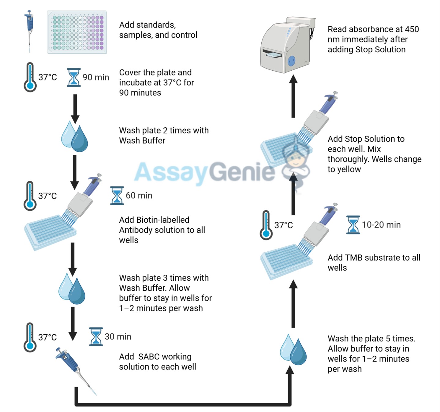
Kit Components

Component Quantity Storage

ELISA Microplate (Dismountable)

8x12 strips

4°C for 6 months

Lyophilized Standard

2

4°C/ -20°C

Sample/Standard Dlution Buffer

20ml

4°C

Biotin-labeled Antibody (Concentrated)

120ul

4°C (Protection from light)

Antibody Dilution Buffer

10ml

4°C

HRP-Streptavidin Conjugate (SABC)

120ul

4°C (Protect from light)

SABC Dilution Buffer

10ml

4°C

TMB Substrate

10ml

4°C (Protection from light)

Stop Solution

10ml

4°C

Wash Buffer (25X)

30ml

4°C

Plate Sealer

5

-

Other materials required:

  • Microplate reader with 450 nm wavelength filter
  • Multichannel Pipette, Pipette, microcentrifuge tubes and disposable pipette tips
  • Incubator
  • Deionized or distilled water
  • Absorbent paper
  • Buffer resevoir

Protein Information

Heading 1 Heading 2

UniProt

NCBI GenInfo Identifier

NCBI Gene ID

Molecular Weight

44,341 Da

Protein Family

Transforming growth factor

Protocol

*Note: Protocols are specific to each batch/lot. For the exact instructions please follow the protocol included in your kit.

Before adding to wells, equilibrate the SABC working solution and TMB substrate for at least 30 min at 37°C. When diluting samples and reagents, they must be mixed completely and evenly. It is recommended to plot a standard curve for each test.

Step Procedure

1.

Set standard, test sample and control (zero) wells on the pre-coated plate respectively, and then, record their positions. It is recommended to measure each standard and sample in duplicate. Wash plate 2 times before adding standard, sample and control (zero) wells!

2.

Aliquot 0.1ml standard solutions into the standard wells.

3.

Add 0.1 ml of Sample / Standard dilution buffer into the control (zero) well.

4.

Add 0.1 ml of properly diluted sample ( Human serum, plasma, tissue homogenates and other biological fluids.) into test sample wells.

5.

Seal the plate with a cover and incubate at 37 °C for 90 min.

6.

Remove the cover and discard the plate content, clap the plate on the absorbent filter papers or other absorbent material. Do NOT let the wells completely dry at any time. Wash plate X2.

7.

Add 0.1 ml of Biotin- detection antibody working solution into the above wells (standard, test sample & zero wells). Add the solution at the bottom of each well without touching the side wall.

8.

Seal the plate with a cover and incubate at 37°C for 60 min.

9.

Remove the cover, and wash plate 3 times with Wash buffer. Let wash buffer rest in wells for 1 min between each wash.

10.

Add 0.1 ml of SABC working solution into each well, cover the plate and incubate at 37°C for 30 min.

11.

Remove the cover and wash plate 5 times with Wash buffer, and each time let the wash buffer stay in the wells for 1-2 min.

12.

Add 90 µl of TMB substrate into each well, cover the plate and incubate at 37°C in dark within 10-20 min. (Note: This incubation time is for reference use only, the optimal time should be determined by end user.) And the shades of blue can be seen in the first 3-4 wells (with most concentrated standard solutions), the other wells show no obvious color.

13.

Add 50 µl of Stop solution into each well and mix thoroughly. The color changes into yellow immediately.

14.

Read the O.D. absorbance at 450 nm in a microplate reader immediately after adding the stop solution.

Sample Type

When carrying out an ELISA assay it is important to prepare your samples in order to achieve the best possible results. Below we have a list of procedures for the preparation of samples for different sample types.

Sample Type Protocol

Serum

If using serum separator tubes, allow samples to clot for 30 minutes at room temperature. Centrifuge for 10 minutes at 1,000x g. Collect the serum fraction and assay promptly or aliquot and store the samples at -80°C. Avoid multiple freeze-thaw cycles.

If serum separator tubes are not being used, allow samples to clot overnight at 2-8°C. Centrifuge for 10 minutes at 1,000x g. Remove serum and assay promptly or aliquot and store the samples at -80°C. Avoid multiple freeze-thaw cycles.

Plasma

Collect plasma using EDTA or heparin as an anticoagulant. Centrifuge samples at 4°C for 15 mins at 1000 × g within 30 mins of collection. Collect the plasma fraction and assay promptly or aliquot and store the samples at -80°C. Avoid multiple freeze-thaw cycles. Note: Over haemolysed samples are not suitable for use with this kit.

Urine & Cerebrospinal Fluid

Collect the urine (mid-stream) in a sterile container, centrifuge for 20 mins at 2000-3000 rpm. Remove supernatant and assay immediately. If any precipitation is detected, repeat the centrifugation step. A similar protocol can be used for cerebrospinal fluid.

Cell culture supernatant

Collect the cell culture media by pipette, followed by centrifugation at 4°C for 20 mins at 1500 rpm. Collect the clear supernatant and assay immediately.

Cell lysates

Solubilize cells in lysis buffer and allow to sit on ice for 30 minutes. Centrifuge tubes at 14,000 x g for 5 minutes to remove insoluble material. Aliquot the supernatant into a new tube and discard the remaining whole cell extract. Quantify total protein concentration using a total protein assay. Assay immediately or aliquot and store at ≤ -20 °C.

Tissue homogenates

The preparation of tissue homogenates will vary depending upon tissue type. Rinse tissue with 1X PBS to remove excess blood & homogenize in 20ml of 1X PBS (including protease inhibitors) and store overnight at ≤ -20°C. Two freeze-thaw cycles are required to break the cell membranes. To further disrupt the cell membranes you can sonicate the samples. Centrifuge homogenates for 5 mins at 5000xg. Remove the supernatant and assay immediately or aliquot and store at -20°C or -80°C.

Tissue lysates

Rinse tissue with PBS, cut into 1-2 mm pieces, and homogenize with a tissue homogenizer in PBS. Add an equal volume of RIPA buffer containing protease inhibitors and lyse tissues at room temperature for 30 minutes with gentle agitation. Centrifuge to remove debris. Quantify total protein concentration using a total protein assay. Assay immediately or aliquot and store at ≤ -20 °C

Breast Milk

Collect milk samples and centrifuge at 10,000 x g for 60 min at 4°C. Aliquot the supernatant and assay. For long term use, store samples at -80°C. Minimize freeze/thaw cycles.

TGF-Beta Background

TGF-beta Background

Transforming growth factor beta (TGF-β) is a highly influential cytokine that plays a pivotal role in diverse physiological and pathological processes within the body. Belonging to a superfamily of proteins with remarkable regulatory functions, TGF-β exhibits multifaceted effects on cellular behavior, including cell growth, differentiation, and immune responses. It is involved in various stages of development, tissue homeostasis, wound healing, and immune system regulation.

TGF-beta Signaling Pathway

The TGF Beta signaling pathway is initiated by binding of TGF-β ligands to specific cell surface receptors, known as TGF-β type I and type II receptors. This ligand-receptor interaction triggers a cascade of intracellular events, leading to the activation of downstream signaling molecules. The canonical TGF Beta pathway involves the phosphorylation of receptor-regulated Smad proteins, which then form complexes with co-Smad proteins and translocate into the nucleus. In the nucleus, these Smad complexes regulate gene expression by interacting with DNA-binding proteins and modulating target gene transcription. In addition to the Smad-dependent pathway, TGF Beta can also activate non-Smad signaling cascades, such as the MAPK and PI3K-AKT pathways, leading to diverse cellular responses. The TGF Beta signaling pathway exerts profound control over cellular behavior, including cell growth, differentiation, apoptosis, immune regulation, and extracellular matrix remodeling, thereby playing a crucial role in development, tissue homeostasis, and disease progression

TGF-Beta ELISA Kit FAQs

What is the TGF-beta ELISA Kit used for?

It enables researchers to investigate the role of TGF-β in various biological processes and disease conditions. By measuring the levels of TGF beta in different samples, researchers can gain insights into the signaling pathways, cellular responses, and regulatory functions associated with TGF-beta.

What are the advantages of using the TGF-beta ELISA Kit?

The TGF-beta ELISA Kit offers several advantages, including high sensitivity, accuracy, and reproducibility. It provides a user-friendly and reliable method to quantify TGF-beta levels in biological specimens, allowing for precise measurements and robust data analysis.

What sample types are compatible with TGF-beta ELISA Kit?

The TGF-beta ELISA Kit is compatible with various sample types, including serum, plasma, cell lysates, and tissue homogenates. It provides flexibility in sample selection, allowing researchers to analyze TGF-beta levels in different biological matrices.

What are the storage requirements with TGF-beta ELISA Kit?

The TGF-beta ELISA Kit components should be stored according to the instructions provided in the kit manual. Generally, it is recommended to store the kit components at the recommended temperature to ensure their stability and optimal performance.

What should I do if my assay results are not optimal?

If you encounter any issues or have suboptimal assay results, we recommend contacting our dedicated support team for assistance. They will be available to provide troubleshooting guidance, answer your questions, and ensure you achieve the best possible results with the TGF-beta ELISA Kit.