The IGHA1 Polyclonal Antibody (CAB8708) is a high-quality antibody developed for reliable detection and analysis of target proteins. This antibody, generated in rabbits, is highly specific to human samples and has been validated for use in Western blot applications. By targeting the Igha1 protein, researchers can accurately detect and analyze its expression in various cell types, making it an essential reagent for studies in immunology, infectious diseases, and autoimmune disorders.The Igha1 protein is a key component of the immune system, playing a crucial role in antibody production and immune defense mechanisms. Understanding its function and regulation is essential for gaining insights into how the immune system responds to pathogens and maintains immune homeostasis.
This antibody is validated for use in WB, FC, ELISA applications and has demonstrated reactivity against Human samples.
Product Name:
IGHA1 Polyclonal Antibody
SKU:
CAB8708
Size:
20μL, 100μL
Reactivity:
Human
Conjugate:
Unconjugated
Immunogen:
Synthetic peptide. This information is considered to be commercially sensitive.
Tested Applications:
WBFCELISA
Recommended Dilution:
WB
1:500 - 1:1000
FC
1:20 - 1:50
ELISA
Recommended starting concentration is 1 μg/mL. Please optimize the concentration based on your specific assay requirements.
Synonyms:
IgA1, IGHA1
Positive Sample:
SH-SY5Y
Cellular Localization:
Blood Microparticle, External Side Of Plasma Membrane, Extracellular Exosome, Extracellular Region, Extracellular Space, Immunoglobulin Complex, Circulating.
Calculated MW:
38kDa
Observed MW:
60kDa
Contributes to immunoglobulin receptor binding activity. Involved in antibacterial humoral response; glomerular filtration; and positive regulation of respiratory burst. Located in extracellular space. Part of monomeric IgA immunoglobulin complex and secretory dimeric IgA immunoglobulin complex.
Purification Method
Affinity purification
Gene ID
3493
RRID
AB_2769932
Buffer Information
Store at -20℃. Avoid freeze / thaw cycles. Buffer: PBS containing 50% glycerol, preserved with proclin300 or sodium azide, pH 7.3.
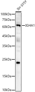
Western blot analysis of lysates from SH-SY5Y cells, using IGHA1 Rabbit pAb (CAB8708) at 1:2000 dilution. Secondary antibody: HRP-conjugated Goat anti-Rabbit IgG (H+L) (CABS014) at 1:10000 dilution. Lysates/proteins: 25μg per lane. Blocking buffer: 3% nonfat dry milk in TBST. Detection: ECL Basic Kit (AbGn00020). Exposure time: 180s.
 at 1:2000 dilution. Secondary antibody: HRP Goat Anti-Rabbit IgG (H+L) at 1:10000 dilution. Lysates/proteins: 25μg per lane. Blocking buffer: 3% nonfat dry milk in TBST.")
 at 1:2000 dilution. Secondary antibody: HRP Goat Anti-Rabbit IgG (H+L) at 1:10000 dilution. Lysates/proteins: 25μg per lane. Blocking buffer: 3% nonfat dry milk in TBST.")

 at 1:10000 dilution. Lysates/proteins: 25ug per lane. Blocking buffer: 3% nonfat dry milk in TBST. Detection: ECL Enhanced Kit. Exposure time: 300s.")

")
. Blue: DAPI for nuclear staining.")
 at 1:10000 dilution. Lysates/proteins: 25ug per lane. Blocking buffer: 3% nonfat dry milk in TBST. Detection: ECL Basic Kit. Exposure time: 180s.")
 variety Zhonghua 11, using OsNAC4 antibody at 1:1000 dilution. Secondary antibody: HRP Goat Anti-Rabbit IgG (H+L) at 1:10000 dilution. Lysates/proteins: 25ug per lane. Blocking buffer: 3% nonfat dry milk in TBST. Detection: ECL Enhanced Kit. Exposure time: 30s.")