The ACAT1 Polyclonal Antibody (CAB5335) is a high-quality antibody developed for reliable detection and analysis of target proteins. This gene encodes a mitochondrially localized enzyme that catalyzes the reversible formation of acetoacetyl-CoA from two molecules of acetyl-CoA. Defects in this gene are associated with 3-ketothiolase deficiency, an inborn error of isoleucine catabolism characterized by urinary excretion of 2-methyl-3-hydroxybutyric acid, 2-methylacetoacetic acid, tiglylglycine, and butanone. RRID AB_2766146 Gene ID 38 Swiss Prot Synonym T2; MAT; ACAT; THIL; ACAT1
This antibody is validated for use in WB, ELISA applications and has demonstrated reactivity against Human, Mouse, Rat samples.
Product Name:
ACAT1 Polyclonal Antibody
SKU:
CAB5335
Size:
100μL, 20μL
Reactivity:
Human, Mouse, Rat
Clone Number:
-
Conjugate:
Unconjugated
Immunogen:
Recombinant protein (or fragment).This information is considered to be commercially sensitive.
Tested Applications:
WBELISA
Recommended Dilution:
WB
1:500 - 1:1000
ELISA
Recommended starting concentration is 1 μg/mL. Please optimize the concentration based on your specific assay requirements.
Synonyms:
T2, MAT, ACAT, THIL, ACAT1
Positive Sample:
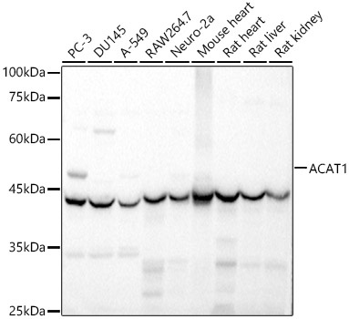
PC-3, DU145, A-549, RAW264.7, Neuro-2a, Mouse heart, Rat heart, Rat liver,Rat kidney
Cellular Localization:
Mitochondrion.
Calculated MW:
45kDa
Observed MW:
42kDa
This gene encodes a mitochondrially localized enzyme that catalyzes the reversible formation of acetoacetyl-CoA from two molecules of acetyl-CoA. Defects in this gene are associated with 3-ketothiolase deficiency, an inborn error of isoleucine catabolism characterized by urinary excretion of 2-methyl-3-hydroxybutyric acid, 2-methylacetoacetic acid, tiglylglycine, and butanone. RRID AB_2766146 Gene ID 38 Swiss Prot Synonym T2; MAT; ACAT; THIL; ACAT1
Purification Method:
Affinity purification
Gene ID:
38
RRID:
AB_2766146
Buffer Information:
Store at -20℃. Avoid freeze / thaw cycles. Buffer: PBS containing 50% glycerol, preserved with proclin300 or sodium azide, pH 7.3.
Western blot analysis of various lysates, using ACAT1 Rabbit pAb (CAB5335) at 1:1000 dilution. Secondary antibody: HRP-conjugated Goat anti-Rabbit IgG (H+L) (AS014) at 1:10000 dilution. Lysates/proteins: 25μg per lane. Blocking buffer: 3% nonfat dry milk in TBST. Detection: ECL Basic Kit (AbGn00020). Exposure time: 1s.


 at 1:10000 dilution. Lysates/proteins: 25ug per lane. Blocking buffer: 3% nonfat dry milk in TBST. Detection: ECL Enhanced Kit. Exposure time: 300s.")

")