The AMPD1 Antibody (CAB3584) is a high-quality antibody developed for reliable detection and analysis of target proteins. Adenosine monophosphate deaminase 1 catalyzes the deamination of AMP to IMP in skeletal muscle and plays an important role in the purine nucleotide cycle. Two other genes have been identified, AMPD2 and AMPD3, for the liver- and erythocyte-specific isoforms, respectively. Deficiency of the muscle-specific enzyme is apparently a common cause of exercise-induced myopathy and probably the most common cause of metabolic myopathy in the human. Alternatively spliced transcript variants encoding different isoforms have been identified in this gene.
This antibody is validated for use in WB, IF/ICC, ELISA applications and has demonstrated reactivity against Human, Mouse, Rat samples.
Product Name:
AMPD1 Antibody
SKU:
CAB3584
Size:
100μL, 20μL
Reactivity:
Human, Mouse, Rat
Conjugate:
Unconjugated
Immunogen:
Recombinant protein (or fragment).This information is considered to be commercially sensitive.
Tested Applications:
WBIF/ICCELISA
Recommended Dilution:
WB
1:500 - 1:2000
IF/ICC
1:50 - 1:200
ELISA
Recommended starting concentration is 1 μg/mL. Please optimize the concentration based on your specific assay requirements.
Synonyms:
MAD, MADA, MMDD, AMPD1
Positive Sample:
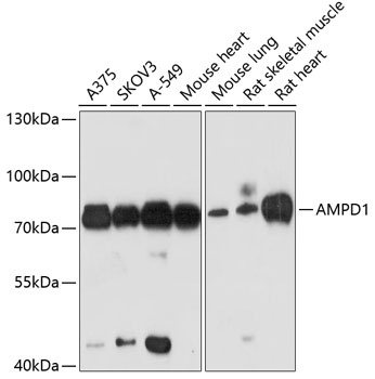
A375, SKOV3, A-549, Mouse heart, Mouse lung, Rat skeletal muscle, Rat heart
Cellular Localization:
Cytosol.
Calculated MW:
86kDa
Observed MW:
80kDa
Adenosine monophosphate deaminase 1 catalyzes the deamination of AMP to IMP in skeletal muscle and plays an important role in the purine nucleotide cycle. Two other genes have been identified, AMPD2 and AMPD3, for the liver- and erythocyte-specific isoforms, respectively. Deficiency of the muscle-specific enzyme is apparently a common cause of exercise-induced myopathy and probably the most common cause of metabolic myopathy in the human. Alternatively spliced transcript variants encoding different isoforms have been identified in this gene.
Purification Method
Affinity purification
Gene ID
270
RRID
AB_2765165
Buffer Information
Store at -20℃. Avoid freeze / thaw cycles. Buffer: PBS containing 50% glycerol, preserved with proclin300 or sodium azide, pH 7.3.
Western blot analysis of various lysates using AMPD1 Rabbit pAb (CAB3584) at 1:3000 dilution. Secondary antibody: HRP-conjugated Goat anti-Rabbit IgG (H+L) (AS014) at 1:10000 dilution. Lysates/proteins: 25μg per lane. Blocking buffer: 3% nonfat dry milk in TBST. Detection: ECL Basic Kit (AbGn00020). Exposure time: 90s.
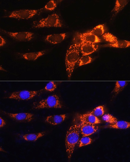
Immunofluorescence analysis of NIH/3T3 cells using AMPD1 Rabbit pAb (CAB3584) at dilution of 1:100. Secondary antibody: Cy3-conjugated Goat anti-Rabbit IgG (H+L) (AS007) at 1:500 dilution. Blue: DAPI for nuclear staining.
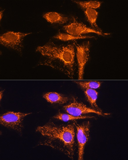
Immunofluorescence analysis of C6 cells using AMPD1 Rabbit pAb (CAB3584) at dilution of 1:100. Secondary antibody: Cy3-conjugated Goat anti-Rabbit IgG (H+L) (AS007) at 1:500 dilution. Blue: DAPI for nuclear staining.