The DNAJC17 Antibody (CAB20168) is a high-quality antibody developed for reliable detection and analysis of target proteins. Predicted to enable RNA binding activity. Predicted to act upstream of or within negative regulation of transcription by RNA polymerase II and toxin transport. Predicted to be located in cytoplasm and nucleus.
This antibody is validated for use in WB, IHC-P, ELISA applications and has demonstrated reactivity against Human, Mouse, Rat samples.
Product Name:
DNAJC17 Antibody
SKU:
CAB20168
Size:
100μL, 20μL
Reactivity:
Human, Mouse, Rat
Conjugate:
Unconjugated
Immunogen:
Recombinant protein (or fragment).This information is considered to be commercially sensitive.
Tested Applications:
WBIHC-PELISA
Recommended Dilution:
WB
1:500 - 1:1000
IHC-P
1:50 - 1:200
ELISA
Recommended starting concentration is 1 μg/mL. Please optimize the concentration based on your specific assay requirements.
Synonyms:
DNAJC17
Positive Sample:
HeLa, A-549, Mouse testis, Mouse thymus, Rat testis
Cellular Localization:
Cytoplasm.
Calculated MW:
35kDa
Observed MW:
35kDa
Predicted to enable RNA binding activity. Predicted to act upstream of or within negative regulation of transcription by RNA polymerase II and toxin transport. Predicted to be located in cytoplasm and nucleus.
Purification Method
Affinity purification
Gene ID
55192
RRID
AB_2862955
Buffer Information
Store at -20℃. Avoid freeze / thaw cycles. Buffer: PBS with 0.01% thimerosal,50% glycerol,pH7.3.
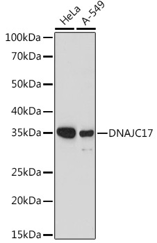
Western blot analysis of various lysates using DNAJC17 Rabbit pAb (CAB20168) at 1:1000 dilution. Secondary antibody: HRP-conjugated Goat anti-Rabbit IgG (H+L) (AS014) at 1:10000 dilution. Lysates/proteins: 25μg per lane. Blocking buffer: 3% nonfat dry milk in TBST. Detection: ECL Basic Kit (AbGn00020). Exposure time: 180s.
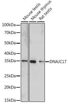
Western blot analysis of various lysates using DNAJC17 Rabbit pAb (CAB20168) at 1:1000 dilution. Secondary antibody: HRP-conjugated Goat anti-Rabbit IgG (H+L) (AS014) at 1:10000 dilution. Lysates/proteins: 25μg per lane. Blocking buffer: 3% nonfat dry milk in TBST. Detection: ECL Enhanced Kit (AbGn00021). Exposure time: 180s.
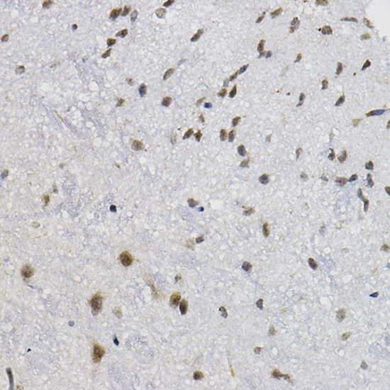
Immunohistochemistry analysis of paraffin-embedded Mouse spinal cord using DNAJC17 Rabbit pAb (CAB20168) at dilution of 1:20 (40x lens). High pressure antigen retrieval performed with 0.01M Citrate buffer (pH 6.0) prior to IHC staining.