The PDIA4 Antibody (CAB4326) is a high-quality antibody developed for reliable detection and analysis of target proteins. This gene encodes a member of the disulfide isomerase (PDI) family of endoplasmic reticulum (ER) proteins that catalyze protein folding and thiol-disulfide interchange reactions. The encoded protein has an N-terminal ER-signal sequence, three catalytically active thioredoxin (TRX) domains, two TRX-like domains and a C-terminal ER-retention sequence. This protein, when bound to cyclophilin B, enhances the rate of immunoglobulin G intermolecular disulfide bonding and antibody assembly.
This antibody is validated for use in WB, IHC-P, IF/ICC, ELISA applications and has demonstrated reactivity against Human, Mouse, Rat samples.
Product Name:
PDIA4 Antibody
SKU:
CAB4326
Size:
100μL, 20μL
Reactivity:
Human, Mouse, Rat
Conjugate:
Unconjugated
Immunogen:
Synthetic peptide. This information is considered to be commercially sensitive.
Tested Applications:
WBIHC-PIF/ICCELISA
Recommended Dilution:
WB
1:500 - 1:1000
IHC-P
1:50 - 1:200
IF/ICC
1:50 - 1:200
ELISA
Recommended starting concentration is 1 μg/mL. Please optimize the concentration based on your specific assay requirements.
Synonyms:
ERP70, ERP72, ERp-72, PDIA4
Positive Sample:
HepG2, MCF7, Mouse liver, Rat lung, Rat liver
Cellular Localization:
Endoplasmic Reticulum Lumen, Melanosome.
Calculated MW:
73kDa
Observed MW:
72kDa
This gene encodes a member of the disulfide isomerase (PDI) family of endoplasmic reticulum (ER) proteins that catalyze protein folding and thiol-disulfide interchange reactions. The encoded protein has an N-terminal ER-signal sequence, three catalytically active thioredoxin (TRX) domains, two TRX-like domains and a C-terminal ER-retention sequence. This protein, when bound to cyclophilin B, enhances the rate of immunoglobulin G intermolecular disulfide bonding and antibody assembly.
Purification Method
Affinity purification
Gene ID
9601
RRID
AB_2765625
Buffer Information
Store at -20℃. Avoid freeze / thaw cycles. Buffer: PBS containing 50% glycerol, preserved with proclin300 or sodium azide, pH 7.3.
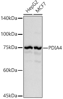
Western blot analysis of various lysates using PDIA4 Rabbit pAb (CAB4326) at 1:1000 dilution. Secondary antibody: HRP-conjugated Goat anti-Rabbit IgG (H+L) (AS014) at 1:10000 dilution. Lysates/proteins: 25μg per lane. Blocking buffer: 3% nonfat dry milk in TBST. Detection: ECL Basic Kit (AbGn00020). Exposure time: 0.5s.
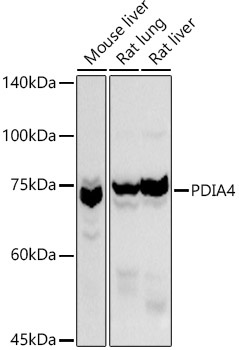
Western blot analysis of various lysates using PDIA4 Rabbit pAb (CAB4326) at 1:1000 dilution. Secondary antibody: HRP-conjugated Goat anti-Rabbit IgG (H+L) (AS014) at 1:10000 dilution. Lysates/proteins: 25μg per lane. Blocking buffer: 3% nonfat dry milk in TBST. Detection: ECL Basic Kit (AbGn00020). Exposure time: 10s.
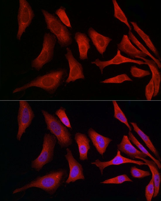
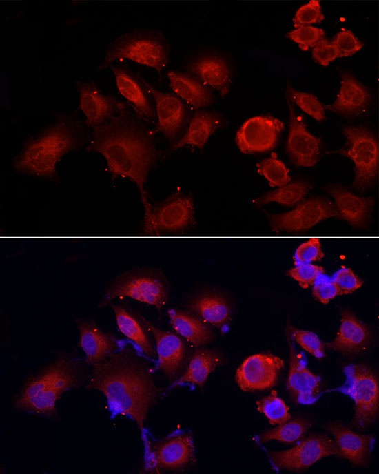
Immunofluorescence analysis of HeLa cells using PDIA4 Rabbit pAb (CAB4326) at dilution of 1:50 (40x lens). Secondary antibody: Cy3-conjugated Goat anti-Rabbit IgG (H+L) (AS007) at 1:500 dilution. Blue: DAPI for nuclear staining.
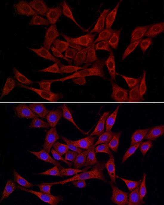
Immunofluorescence analysis of NIH/3T3 cells using PDIA4 Rabbit pAb (CAB4326) at dilution of 1:50 (40x lens). Secondary antibody: Cy3-conjugated Goat anti-Rabbit IgG (H+L) (AS007) at 1:500 dilution. Blue: DAPI for nuclear staining.
Immunofluorescence analysis of PC-3 cells using PDIA4 Rabbit pAb (CAB4326) at dilution of 1:50 (40x lens). Secondary antibody: Cy3-conjugated Goat anti-Rabbit IgG (H+L) (AS007) at 1:500 dilution. Blue: DAPI for nuclear staining.