The ASPSCR1 Antibody (CAB7481) is a high-quality antibody developed for reliable detection and analysis of target proteins. The protein encoded by this gene contains a UBX domain and interacts with glucose transporter type 4 (GLUT4). This protein is a tether, which sequesters the GLUT4 in intracellular vesicles in muscle and fat cells in the absence of insulin, and redistributes the GLUT4 to the plasma membrane within minutes of insulin stimulation. Translocation t(X;17)(p11;q25) of this gene with transcription factor TFE3 gene results in a ASPSCR1-TFE3 fusion protein in alveolar soft part sarcoma and in renal cell carcinomas. Multiple alternatively spliced transcript variants have been found.
This antibody is validated for use in WB, IF/ICC, IP, ELISA applications and has demonstrated reactivity against Human, Mouse, Rat samples.
Product Name:
ASPSCR1 Antibody
SKU:
CAB7481
Size:
100μL, 20μL
Reactivity:
Human, Mouse, Rat
Conjugate:
Unconjugated
Immunogen:
Recombinant protein (or fragment).This information is considered to be commercially sensitive.
Tested Applications:
WBIF/ICCIPELISA
Recommended Dilution:
WB
1:500 - 1:2000
IF/ICC
1:50 - 1:200
IP
0.5μg-4μg antibody for 200μg-400μg extracts of whole cells
ELISA
Recommended starting concentration is 1 μg/mL. Please optimize the concentration based on your specific assay requirements.
The protein encoded by this gene contains a UBX domain and interacts with glucose transporter type 4 (GLUT4). This protein is a tether, which sequesters the GLUT4 in intracellular vesicles in muscle and fat cells in the absence of insulin, and redistributes the GLUT4 to the plasma membrane within minutes of insulin stimulation. Translocation t(X;17)(p11;q25) of this gene with transcription factor TFE3 gene results in a ASPSCR1-TFE3 fusion protein in alveolar soft part sarcoma and in renal cell carcinomas. Multiple alternatively spliced transcript variants have been found.
Purification Method
Affinity purification
Gene ID
79058
RRID
AB_2768012
Buffer Information
Store at -20℃. Avoid freeze / thaw cycles. Buffer: PBS containing 50% glycerol, preserved with proclin300 or sodium azide, pH 7.3.
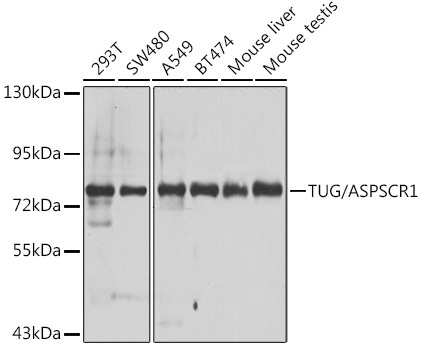
Western blot analysis of various lysates using TUG/ASPSCR1 Rabbit pAb (CAB7481) at 1:1000 dilution. Secondary antibody: HRP-conjugated Goat anti-Rabbit IgG (H+L) (AS014) at 1:10000 dilution. Lysates/proteins: 25μg per lane. Blocking buffer: 3% nonfat dry milk in TBST. Detection: ECL Basic Kit (AbGn00020). Exposure time: 30s.
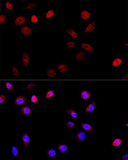
Immunofluorescence analysis of HeLa cells using TUG/ASPSCR1 Rabbit pAb (CAB7481) at dilution of 1:100 (40x lens). Secondary antibody: Cy3-conjugated Goat anti-Rabbit IgG (H+L) (AS007) at 1:500 dilution. Blue: DAPI for nuclear staining.
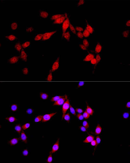
Immunofluorescence analysis of PC-12 cells using TUG/ASPSCR1 Rabbit pAb (CAB7481) at dilution of 1:100 (40x lens). Secondary antibody: Cy3-conjugated Goat anti-Rabbit IgG (H+L) (AS007) at 1:500 dilution. Blue: DAPI for nuclear staining.
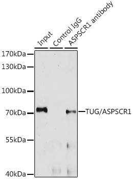
Immunoprecipitation analysis of 200 μg extracts of A-549 cells, using 3 μg TUG/ASPSCR1 antibody (CAB7481). Western blot was performed from the immunoprecipitate using TUG/ASPSCR1 antibody (CAB7481) at a dilution of 1:1000.