The Bcl-2 Monoclonal Antibody (CAB19693) is a high-quality antibody developed for reliable detection and analysis of target proteins. This gene encodes an integral outer mitochondrial membrane protein that blocks the apoptotic death of some cells such as lymphocytes. Constitutive expression of BCL2, such as in the case of translocation of BCL2 to Ig heavy chain locus, is thought to be the cause of follicular lymphoma. Alternative splicing results in multiple transcript variants. RRID AB_2862738 Gene ID 596 Swiss Prot Synonym Bcl-2; PPP1R50
This antibody is validated for use in WB, IHC-P, IP, ELISA applications and has demonstrated reactivity against Human, Mouse samples.
Product Name:
Bcl-2 Monoclonal Antibody
SKU:
CAB19693
Size:
100μL, 20μL
Reactivity:
Human, Mouse
Clone Number:
ARC0173
Conjugate:
Unconjugated
Immunogen:
Recombinant protein (or fragment).This information is considered to be commercially sensitive.
Tested Applications:
WBIHC-PIPELISA
Recommended Dilution:
WB
1:3000 - 1:15000
IHC-P
1:1000 - 1:4000
IP
0.5μg-4μg antibody for 100μg-300μg extracts of whole cells
ELISA
Recommended starting concentration is 1 μg/mL. Please optimize the concentration based on your specific assay requirements.
This gene encodes an integral outer mitochondrial membrane protein that blocks the apoptotic death of some cells such as lymphocytes. Constitutive expression of BCL2, such as in the case of translocation of BCL2 to Ig heavy chain locus, is thought to be the cause of follicular lymphoma. Alternative splicing results in multiple transcript variants. RRID AB_2862738 Gene ID 596 Swiss Prot Synonym Bcl-2; PPP1R50
Purification Method:
Affinity purification
Gene ID:
596
RRID:
AB_2862738
Buffer Information:
Store at -20℃. Avoid freeze / thaw cycles. Buffer: PBS containing 50% glycerol and 0.05% BSA, preserved with proclin300 or sodium azide, pH 7.3.
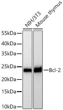
Western blot analysis of various lysates using Bcl-2 Rabbit mAb (CAB19693) at 1:7000 dilution incubated overnight at 4℃. Secondary antibody: HRP-conjugated Goat anti-Rabbit IgG (H+L) (AS014) at 1:10000 dilution. Lysates/proteins: 25 μg per lane. Blocking buffer: 3% nonfat dry milk in TBST. Detection: ECL Basic Kit (AbGn00020). Exposure time: 45 s.
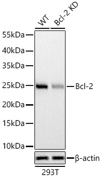
Western blot analysis of lysates from wild type (WT) and Bcl-2 knockdown (KD) 293T cells using Bcl-2 Rabbit mAb (CAB19693) at 1:7000 dilution incubated overnight at 4℃. Secondary antibody: HRP-conjugated Goat anti-Rabbit IgG (H+L) (AS014) at 1:10000 dilution. Lysates/proteins: 25 μg per lane. Blocking buffer: 3% nonfat dry milk in TBST. Detection: ECL Basic Kit (AbGn00020). Exposure time: 90 s.
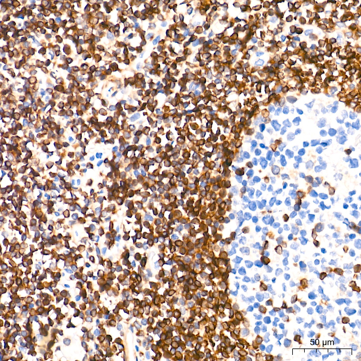
Immunohistochemistry analysis of paraffin-embedded Human tonsil tissue using Bcl-2 Rabbit mAb (CAB19693) at a dilution of 1:2000 (40x lens). High pressure antigen retrieval performed with 0.01M Tris-EDTA Buffer (pH 9.0) prior to IHC staining.
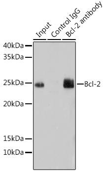
Immunoprecipitation analysis of 200 μg extracts of Mouse lung using 3 μg Bcl-2 Rabbit mAb (CAB19693). Western blot was performed from the immunoprecipitate using Bcl-2 Rabbit mAb (CAB19693) at a dilution of 1:1000.