The BCL3 Antibody (CAB11582) is a high-quality antibody developed for reliable detection and analysis of target proteins. This gene is a proto-oncogene candidate. It is identified by its translocation into the immunoglobulin alpha-locus in some cases of B-cell leukemia. The protein encoded by this gene contains seven ankyrin repeats, which are most closely related to those found in I kappa B proteins. This protein functions as a transcriptional co-activator that activates through its association with NF-kappa B homodimers. The expression of this gene can be induced by NF-kappa B, which forms a part of the autoregulatory loop that controls the nuclear residence of p50 NF-kappa B.
This antibody is validated for use in WB, IF/ICC, ELISA applications and has demonstrated reactivity against Human, Mouse, Rat samples.
Product Name:
BCL3 Antibody
SKU:
CAB11582
Size:
100μL, 20μL
Reactivity:
Human, Mouse, Rat
Conjugate:
Unconjugated
Immunogen:
Synthetic peptide. This information is considered to be commercially sensitive.
Tested Applications:
WBIF/ICCELISA
Recommended Dilution:
WB
1:500 - 1:1000
IF/ICC
1:50 - 1:200
ELISA
Recommended starting concentration is 1 μg/mL. Please optimize the concentration based on your specific assay requirements.
Synonyms:
BCL4, D19S37, BCL3
Positive Sample:
Raji, 293T
Cellular Localization:
Cytoplasm, Nucleus, Perinuclear Region.
Calculated MW:
48kDa
Observed MW:
60kDa
This gene is a proto-oncogene candidate. It is identified by its translocation into the immunoglobulin alpha-locus in some cases of B-cell leukemia. The protein encoded by this gene contains seven ankyrin repeats, which are most closely related to those found in I kappa B proteins. This protein functions as a transcriptional co-activator that activates through its association with NF-kappa B homodimers. The expression of this gene can be induced by NF-kappa B, which forms a part of the autoregulatory loop that controls the nuclear residence of p50 NF-kappa B.
Purification Method
Affinity purification
Gene ID
602
RRID
AB_2758612
Buffer Information
Store at -20℃. Avoid freeze / thaw cycles. Buffer: PBS with 0.01% thimerosal,50% glycerol,pH7.3.
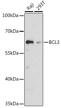
Western blot analysis of various lysates using BCL3 Rabbit pAb (CAB11582) at 1:1000 dilution. Secondary antibody: HRP-conjugated Goat anti-Rabbit IgG (H+L) (AS014) at 1:10000 dilution. Lysates/proteins: 25μg per lane. Blocking buffer: 3% nonfat dry milk in TBST. Detection: ECL Basic Kit (AbGn00020). Exposure time: 30s.
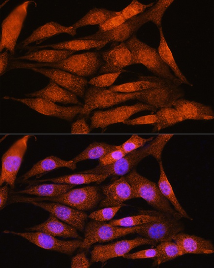
Immunofluorescence analysis of NIH/3T3 cells using BCL3 Rabbit pAb (CAB11582) at dilution of 1:100. Secondary antibody: Cy3-conjugated Goat anti-Rabbit IgG (H+L) (AS007) at 1:500 dilution. Blue: DAPI for nuclear staining.
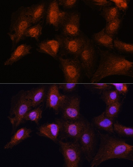
Immunofluorescence analysis of C6 cells using BCL3 Rabbit pAb (CAB11582) at dilution of 1:100. Secondary antibody: Cy3-conjugated Goat anti-Rabbit IgG (H+L) (AS007) at 1:500 dilution. Blue: DAPI for nuclear staining.