The beta-Actin Monoclonal Antibody (High Dilution) (CABC026) is a high-quality antibody developed for reliable detection and analysis of target proteins. This gene encodes one of six different actin proteins. Actins are highly conserved proteins that are involved in cell motility, structure, integrity, and intercellular signaling. The encoded protein is a major constituent of the contractile apparatus and one of the two nonmuscle cytoskeletal actins that are ubiquitously expressed. Mutations in this gene cause Baraitser-Winter syndrome 1, which is characterized by intellectual disability with a distinctive facial appearance in human patients. Numerous pseudogenes of this gene have been identified throughout the human genome. Note:Due to the high antibody titer, it is advisable to be diluted before use. Please dilute 5μL of the antibody solution with 45μL of PBS solution, containing 50% glycerol. The diluted antibody can be stored at -20°C.
This antibody is validated for use in WB, IHC-P, IF/ICC, ELISA applications and has demonstrated reactivity against Human, Mouse, Rat, Chicken, Zebrafish, Pig, Cow samples.
Product Name:
beta-Actin Monoclonal Antibody (High Dilution)
SKU:
CABC026
Size:
100μL, 200μL, 50μL
Reactivity:
Human, Mouse, Rat, Chicken, Zebrafish, Pig, Cow
Clone Number:
ARC5115-01
Conjugate:
Unconjugated
Immunogen:
Recombinant protein (or fragment).This information is considered to be commercially sensitive.
This gene encodes one of six different actin proteins. Actins are highly conserved proteins that are involved in cell motility, structure, integrity, and intercellular signaling. The encoded protein is a major constituent of the contractile apparatus and one of the two nonmuscle cytoskeletal actins that are ubiquitously expressed. Mutations in this gene cause Baraitser-Winter syndrome 1, which is characterized by intellectual disability with a distinctive facial appearance in human patients. Numerous pseudogenes of this gene have been identified throughout the human genome. Note:Due to the high antibody titer, it is advisable to be diluted before use. Please dilute 5μL of the antibody solution with 45μL of PBS solution, containing 50% glycerol. The diluted antibody can be stored at -20°C.
Purification Method
Affinity purification
Gene ID
60
RRID
AB_2768234
Buffer Information
Store at -20℃. Avoid freeze / thaw cycles. Buffer: PBS containing 50% glycerol and 0.05% BSA, preserved with proclin300 or sodium azide, pH 7.3.
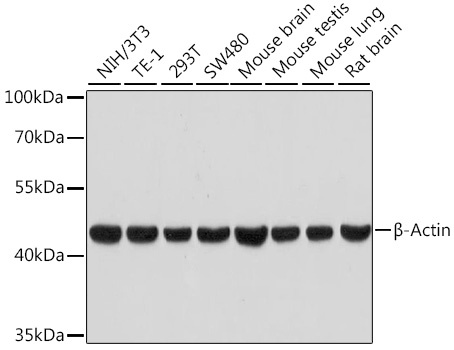
Western blot analysis of various lysates using β-Actin Rabbit mAb (High Dilution) (CABC026) at 1:100000 dilution. Secondary antibody: HRP-conjugated Goat anti-Rabbit IgG (H+L) (AS014) at 1:10000 dilution. Lysates/proteins: 25μg per lane. Blocking buffer: 3% nonfat dry milk in TBST. Detection: ECL Basic Kit (AbGn00020). Exposure time: 30s.
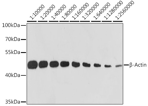
Western blot analysis of lysates from HeLa cells, using β-Actin Rabbit mAb (High Dilution) (CABC026) at 1:10000-1:2560000 dilution. Secondary antibody: HRP-conjugated Goat anti-Rabbit IgG (H+L) (AS014) at 1:10000 dilution. Lysates/proteins: 25μg per lane. Blocking buffer: 3% nonfat dry milk in TBST. Detection: ECL Basic Kit (AbGn00020). Exposure time: 10s.
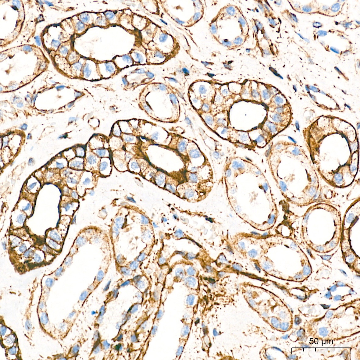
Immunohistochemistry analysis of paraffin-embedded Human kidney tissue using β-Actin Rabbit mAb (High Dilution) (CABC026) at a dilution of 1:10000 (40x lens). High pressure antigen retrieval performed with 0.01M Tris-EDTA Buffer (pH 9.0) prior to IHC staining.
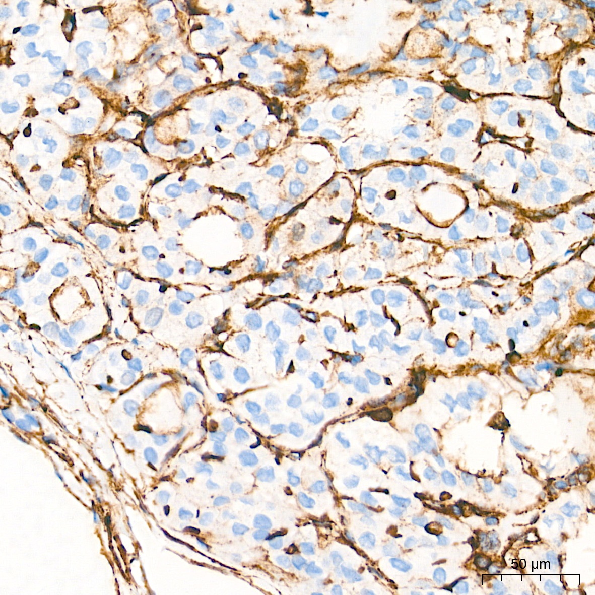
Immunohistochemistry analysis of paraffin-embedded Human thyroid cancer tissue using β-Actin Rabbit mAb (High Dilution) (CABC026) at a dilution of 1:10000 (40x lens). High pressure antigen retrieval performed with 0.01M Tris-EDTA Buffer (pH 9.0) prior to IHC staining.
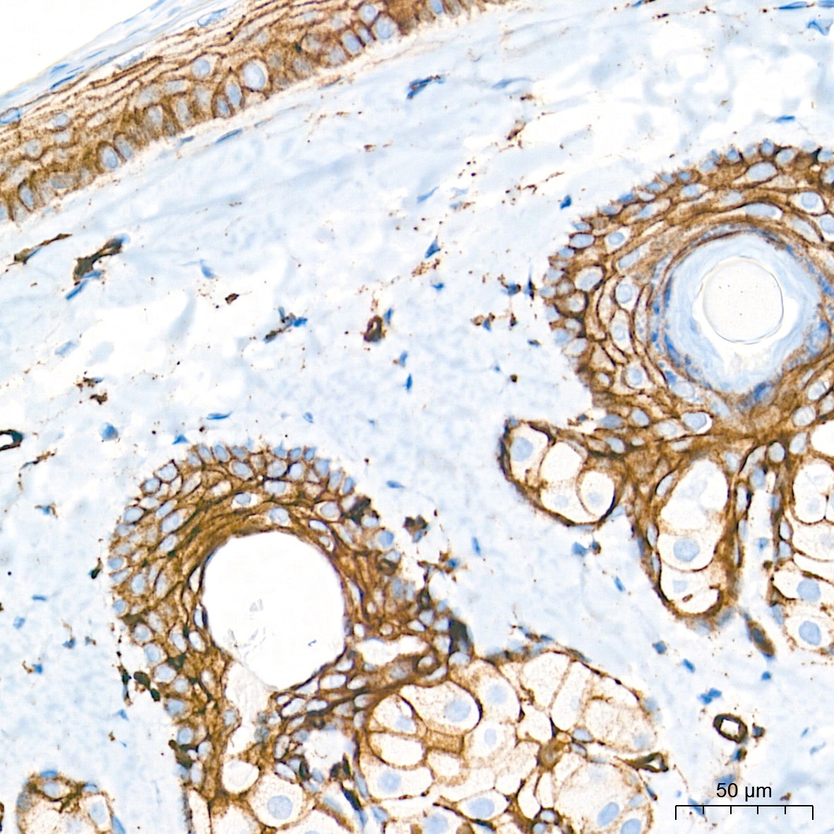
Immunohistochemistry analysis of paraffin-embedded Rat skin tissue using β-Actin Rabbit mAb (High Dilution) (CABC026) at a dilution of 1:10000 (40x lens). High pressure antigen retrieval performed with 0.01M Tris-EDTA Buffer (pH 9.0) prior to IHC staining.
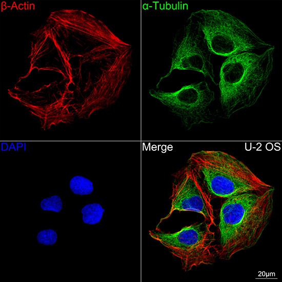
Confocal imaging of U-2 OS cells using β-Actin Rabbit mAb (High Dilution) (CABC026, dilution 1:500) followed by a further incubation with Cy3 Goat Anti-Rabbit IgG (H+L) (AS007, dilution 1:500) (Red). The cells were counterstained with α-Tubulin Mouse mAb (AC012, dilution 1:400) followed by incubation with ABflo® 488-conjugated Goat Anti-Mouse IgG (H+L) Ab (AS076, dilution 1:500) (Green). DAPI was used for nuclear staining (Blue). Objective: 100x.
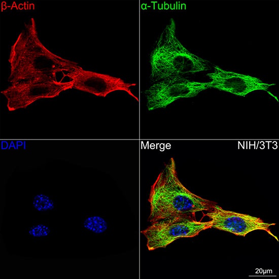
Confocal imaging of NIH/3T3 cells using β-Actin Rabbit mAb (High Dilution) (CABC026, dilution 1:500) followed by a further incubation with Cy3 Goat Anti-Rabbit IgG (H+L) (AS007, dilution 1:500) (Red). The cells were counterstained with α-Tubulin Mouse mAb (AC012, dilution 1:400) followed by incubation with ABflo® 488-conjugated Goat Anti-Mouse IgG (H+L) Ab (AS076, dilution 1:500) (Green). DAPI was used for nuclear staining (Blue). Objective: 100x.
 (CABC026)")
 (CABC026)")
")


