The CAB39 Antibody (CAB16125) is a high-quality antibody developed for reliable detection and analysis of target proteins. Enables kinase binding activity and protein serine/threonine kinase activator activity. Involved in intracellular signal transduction; peptidyl-serine phosphorylation; and positive regulation of protein phosphorylation. Located in extracellular exosome. Implicated in hepatocellular carcinoma. Biomarker of hepatocellular carcinoma and pancreatic cancer. RRID AB_2763570 Gene ID 51719 Swiss Prot Synonym MO25; CGI-66; CAB39
This antibody is validated for use in WB, IF/ICC, ELISA applications and has demonstrated reactivity against Human, Mouse, Rat samples.
Product Name:
CAB39 Antibody
SKU:
CAB16125
Size:
100μL, 20μL
Reactivity:
Human, Mouse, Rat
Clone Number:
-
Conjugate:
Unconjugated
Immunogen:
Synthetic peptide. This information is considered to be commercially sensitive.
Tested Applications:
WBIF/ICCELISA
Recommended Dilution:
WB
1:500 - 1:1000
IF
/
ICC
1:50 - 1:200
ELISA
Recommended starting concentration is 1 μg/mL. Please optimize the concentration based on your specific assay requirements.
Synonyms:
MO25, CGI-66, CAB39
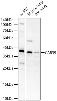
Positive Sample:
K-562, Mouse lung, Rat lung
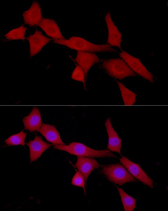
Cellular Localization:
Cytoplasm.
Calculated MW:
40kDa
Observed MW:
39kDa
Enables kinase binding activity and protein serine/threonine kinase activator activity. Involved in intracellular signal transduction; peptidyl-serine phosphorylation; and positive regulation of protein phosphorylation. Located in extracellular exosome. Implicated in hepatocellular carcinoma. Biomarker of hepatocellular carcinoma and pancreatic cancer. RRID AB_2763570 Gene ID 51719 Swiss Prot Synonym MO25; CGI-66; CAB39
Purification Method:
Affinity purification
Gene ID:
51719
RRID:
AB_2763570
Buffer Information:
Store at -20℃. Avoid freeze / thaw cycles. Buffer: PBS containing 50% glycerol, preserved with proclin300 or sodium azide, pH 7.3.
Western blot analysis of various lysates, using CAB39 Rabbit pAb (CAB16125) at 1:700 dilution. Secondary antibody: HRP-conjugated Goat anti-Rabbit IgG (H+L) (AS014) at 1:10000 dilution. Lysates/proteins: 25μg per lane. Blocking buffer: 3% nonfat dry milk in TBST. Detection: ECL Basic Kit (AbGn00020). Exposure time: 60s.
Immunofluorescence analysis of NIH/3T3 cells using CAB39 Rabbit pAb (CAB16125) at dilution of 1:100 (40x lens). Secondary antibody: Cy3-conjugated Goat anti-Rabbit IgG (H+L) (AS007) at 1:500 dilution. Blue: DAPI for nuclear staining.