The Calreticulin Polyclonal Antibody (CAB1066) is a high-quality antibody developed for reliable detection and analysis of target proteins. Calreticulin is a highly conserved chaperone protein which resides primarily in the endoplasmic reticulum, and is involved in a variety of cellular processes, among them, cell adhesion. Additionally, it functions in protein folding quality control and calcium homeostasis. Calreticulin is also found in the nucleus, suggesting that it may have a role in transcription regulation. Systemic lupus erythematosus is associated with increased autoantibody titers against calreticulin. Recurrent mutations in calreticulin have been linked to various neoplasms, including the myeloproliferative type.
This antibody is validated for use in WB, IHC-P, IF/ICC, ELISA applications and has demonstrated reactivity against Human, Mouse, Rat samples.
Product Name:
Calreticulin Polyclonal Antibody
SKU:
CAB1066
Size:
100μL, 20μL
Reactivity:
Human, Mouse, Rat
Conjugate:
Unconjugated
Immunogen:
Recombinant protein (or fragment).This information is considered to be commercially sensitive.
Tested Applications:
WBIHC-PIF/ICCELISA
Recommended Dilution:
WB
1:1000 - 1:5000
IHC-P
1:50 - 1:200
IF/ICC
1:50 - 1:200
ELISA
Recommended starting concentration is 1 μg/mL. Please optimize the concentration based on your specific assay requirements.
Synonyms:
RO, CRT, SSA, cC1qR, HEL-S-99n, Calreticulin
Positive Sample:
HL-60, SH-SY5Y, HeLa, PC-3, C6, SH-SY5Y, mouse kidney, rat liver, rat testis
Calreticulin is a highly conserved chaperone protein which resides primarily in the endoplasmic reticulum, and is involved in a variety of cellular processes, among them, cell adhesion. Additionally, it functions in protein folding quality control and calcium homeostasis. Calreticulin is also found in the nucleus, suggesting that it may have a role in transcription regulation. Systemic lupus erythematosus is associated with increased autoantibody titers against calreticulin. Recurrent mutations in calreticulin have been linked to various neoplasms, including the myeloproliferative type.
Purification Method
Affinity purification
Gene ID
811
RRID
AB_2758161
Buffer Information
Store at -20℃. Avoid freeze / thaw cycles. Buffer: PBS containing 50% glycerol, preserved with proclin300 or sodium azide, pH 7.3.
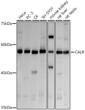
Western blot analysis of various lysates using Calreticulin Rabbit pAb (CAB1066) at 1:3000 dilution. Secondary antibody: HRP-conjugated Goat anti-Rabbit IgG (H+L) (AS014) at 1:10000 dilution. Lysates/proteins: 25μg per lane. Blocking buffer: 3% nonfat dry milk in TBST. Detection: ECL Basic Kit (AbGn00020). Exposure time: 1s.
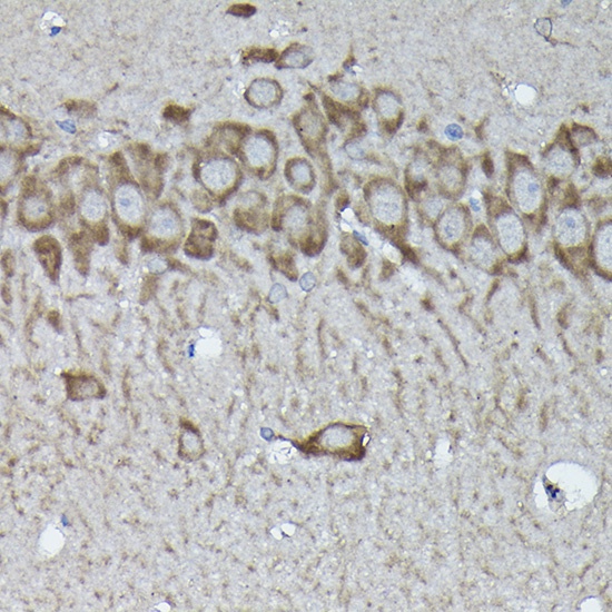
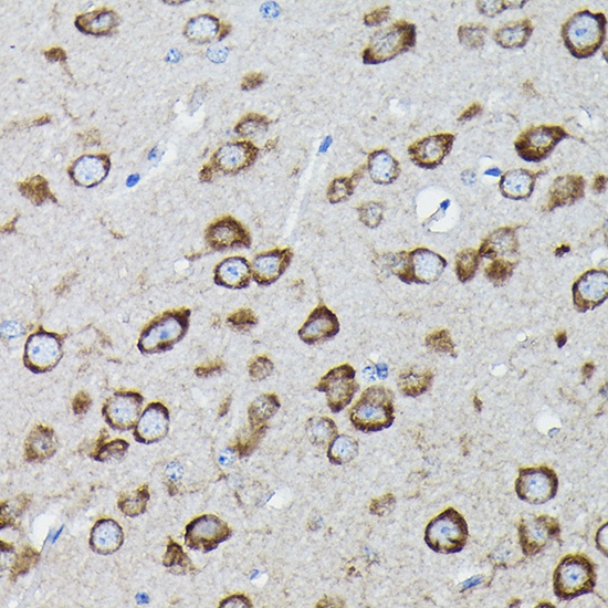
Immunohistochemistry analysis of paraffin-embedded Rat brain using Calreticulineticulin Rabbit pAb (CAB1066) at dilution of 1:100 (40x lens). High pressure antigen retrieval performed with 0.01M Citrate buffer (pH 6.0) prior to IHC staining.
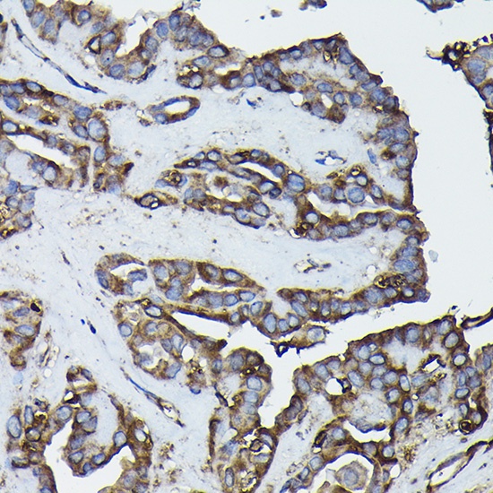
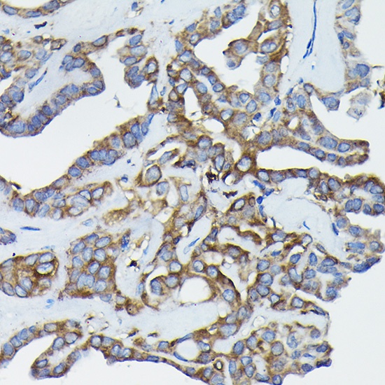
Immunohistochemistry analysis of paraffin-embedded Human thyroid cancer using Calreticulineticulin Rabbit pAb (CAB1066) at dilution of 1:100 (40x lens). High pressure antigen retrieval performed with 0.01M Citrate buffer (pH 6.0) prior to IHC staining.
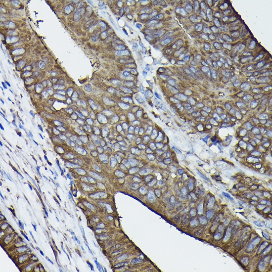
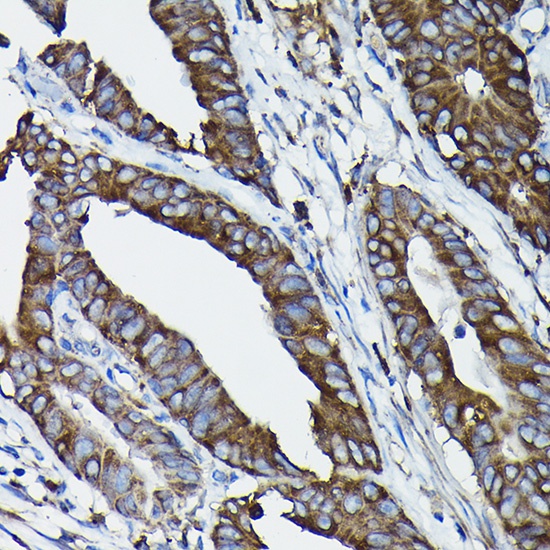
Immunohistochemistry analysis of paraffin-embedded Human colon carcinoma using Calreticulineticulin Rabbit pAb (CAB1066) at dilution of 1:100 (40x lens). High pressure antigen retrieval performed with 0.01M Citrate buffer (pH 6.0) prior to IHC staining.
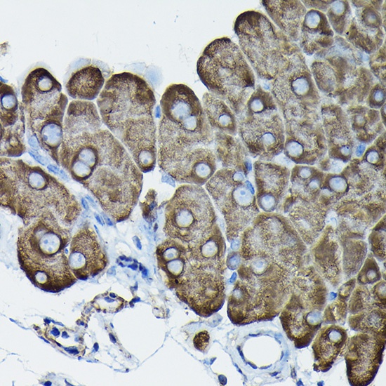
Immunohistochemistry analysis of paraffin-embedded Mouse pancreas using Calreticulineticulin Rabbit pAb (CAB1066) at dilution of 1:100 (40x lens). High pressure antigen retrieval performed with 0.01M Citrate buffer (pH 6.0) prior to IHC staining.
Immunohistochemistry analysis of paraffin-embedded Human colon carcinoma tissue using Calreticulin Rabbit pAb (CAB1066) at a dilution of 1:100 (40x lens). High pressure antigen retrieval was performed with 0.01 M citrate buffer (pH 6.0) prior to IHC staining.
Immunohistochemistry analysis of paraffin-embedded Human thyroid cancer tissue using Calreticulin Rabbit pAb (CAB1066) at a dilution of 1:100 (40x lens). High pressure antigen retrieval was performed with 0.01 M citrate buffer (pH 6.0) prior to IHC staining.
Immunohistochemistry analysis of paraffin-embedded Mouse brain tissue using Calreticulin Rabbit pAb (CAB1066) at a dilution of 1:100 (40x lens). High pressure antigen retrieval was performed with 0.01 M citrate buffer (pH 6.0) prior to IHC staining.
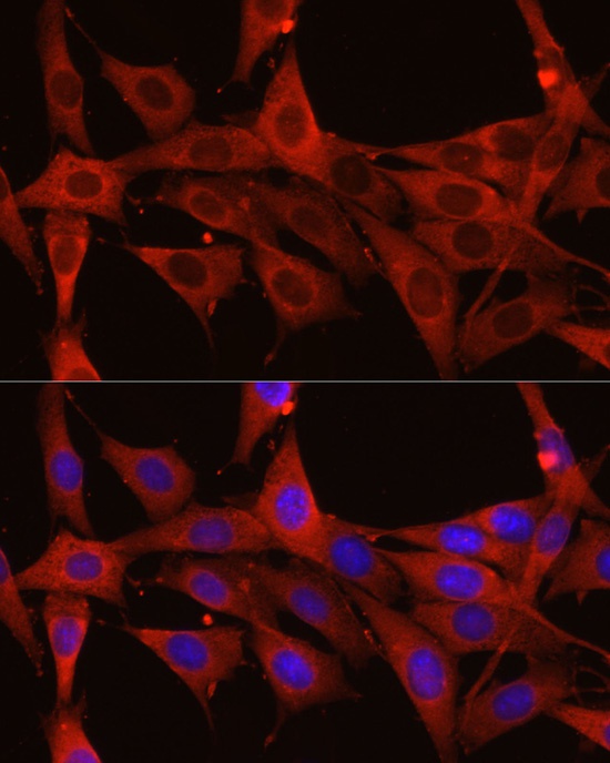
Immunofluorescence analysis of NIH/3T3 cells using Calreticulin Rabbit pAb (CAB1066) at dilution of 1:200 (40x lens). Secondary antibody: Cy3-conjugated Goat anti-Rabbit IgG (H+L) (AS007) at 1:500 dilution. Blue: DAPI for nuclear staining.
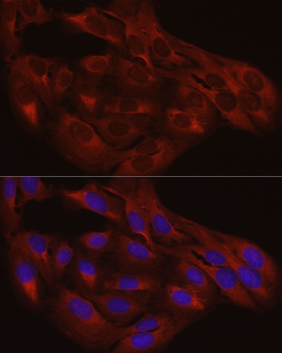
Immunofluorescence analysis of U2OS cells using Calreticulin Rabbit pAb (CAB1066) at dilution of 1:200 (40x lens). Secondary antibody: Cy3-conjugated Goat anti-Rabbit IgG (H+L) (AS007) at 1:500 dilution. Blue: DAPI for nuclear staining.