The Caspase-7 Antibody (CAB1524) is a high-quality antibody developed for reliable detection and analysis of target proteins. This gene encodes a member of the cysteine-aspartic acid protease (caspase) family. Sequential activation of caspases plays a central role in the execution-phase of cell apoptosis. Caspases exist as inactive proenzymes which undergo proteolytic processing at conserved aspartic residues to produce two subunits, large and small, that dimerize to form the active enzyme. The precursor of the encoded protein is cleaved by caspase 3 and 10, is activated upon cell death stimuli and induces apoptosis. Alternatively spliced transcript variants encoding multiple isoforms have been observed for this gene. RRID AB_2762135 Gene ID 840 Swiss Prot Synonym MCH3; CMH-1; LICE2; CASP-7; ICE-LAP3; Caspase-7
This antibody is validated for use in WB, IHC-P, IF/ICC, ELISA applications and has demonstrated reactivity against Human, Mouse, Rat samples.
Product Name:
Caspase-7 Antibody
SKU:
CAB1524
Size:
100μL, 20μL
Reactivity:
Human, Mouse, Rat
Clone Number:
-
Conjugate:
Unconjugated
Immunogen:
Synthetic peptide. This information is considered to be commercially sensitive.
Tested Applications:
WBIHC-PIF/ICCELISA
Recommended Dilution:
WB
1:500 - 1:1000
IHC-P
1:50 - 1:200
IF
/
ICC
1:50 - 1:200
ELISA
Recommended starting concentration is 1 μg/mL. Please optimize the concentration based on your specific assay requirements.
Synonyms:
MCH3, CMH-1, LICE2, CASP-7, ICE-LAP3, Caspase-7
Positive Sample:
Mouse liver, Rat liver
Cellular Localization:
Cytoplasm.
Calculated MW:
34kDa
Observed MW:
35kDa
This gene encodes a member of the cysteine-aspartic acid protease (caspase) family. Sequential activation of caspases plays a central role in the execution-phase of cell apoptosis. Caspases exist as inactive proenzymes which undergo proteolytic processing at conserved aspartic residues to produce two subunits, large and small, that dimerize to form the active enzyme. The precursor of the encoded protein is cleaved by caspase 3 and 10, is activated upon cell death stimuli and induces apoptosis. Alternatively spliced transcript variants encoding multiple isoforms have been observed for this gene. RRID AB_2762135 Gene ID 840 Swiss Prot Synonym MCH3; CMH-1; LICE2; CASP-7; ICE-LAP3; Caspase-7
Purification Method:
Affinity purification
Gene ID:
840
RRID:
AB_2762135
Buffer Information:
Store at -20℃. Avoid freeze / thaw cycles. Buffer: PBS containing 50% glycerol, preserved with proclin300 or sodium azide, pH 7.3.
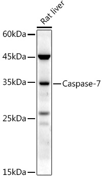
Western blot analysis of lysates from Rat liver, using Caspase-7 Rabbit pAb (CAB1524) at 1:500 dilution. Secondary antibody: HRP-conjugated Goat anti-Rabbit IgG (H+L) (AS014) at 1:10000 dilution. Lysates/proteins: 25μg per lane. Blocking buffer: 3% nonfat dry milk in TBST. Detection: ECL Basic Kit (AbGn00020). Exposure time: 90s.
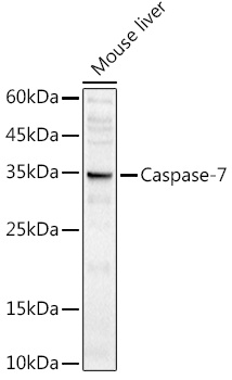
Western blot analysis of lysates from Mouse liver, using Caspase-7 Rabbit pAb (CAB1524) at 1:1000 dilution. Secondary antibody: HRP-conjugated Goat anti-Rabbit IgG (H+L) (AS014) at 1:10000 dilution. Lysates/proteins: 25μg per lane. Blocking buffer: 3% nonfat dry milk in TBST. Detection: ECL Basic Kit (AbGn00020). Exposure time: 3s.
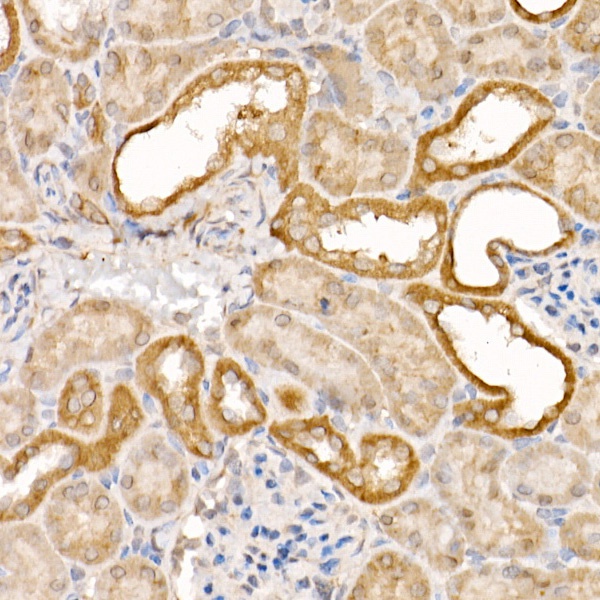
Immunohistochemistry analysis of paraffin-embedded Mouse kidney using Caspase-7 Rabbit pAb (CAB1524) at dilution of 1:100 (40x lens). High pressure antigen retrieval performed with 0.01M Citrate buffer (pH 6.0) prior to IHC staining.
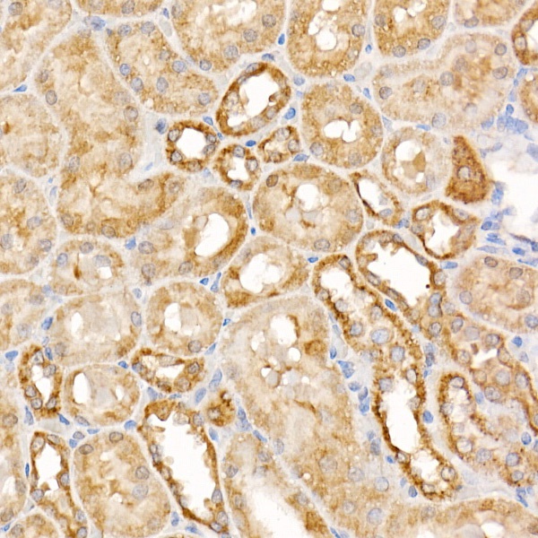
Immunohistochemistry analysis of paraffin-embedded Rat kidney using Caspase-7 Rabbit pAb (CAB1524) at dilution of 1:100 (40x lens). High pressure antigen retrieval performed with 0.01M Citrate buffer (pH 6.0) prior to IHC staining.
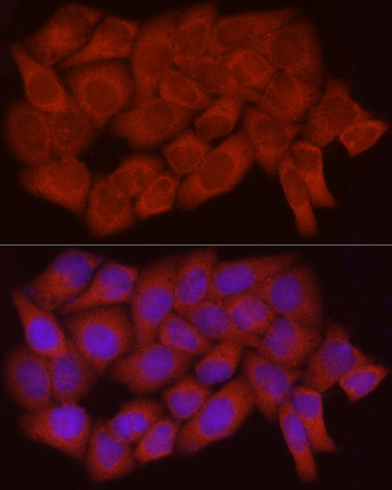
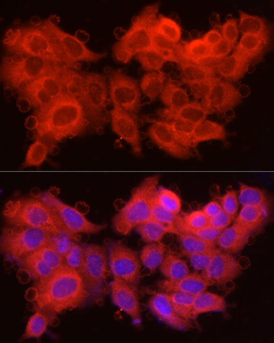
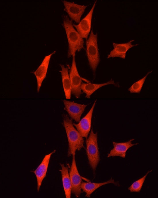
Immunofluorescence analysis of HeLa cells using Caspase-7 Rabbit pAb (CAB1524) at dilution of 1:200 (40x lens). Secondary antibody: Cy3-conjugated Goat anti-Rabbit IgG (H+L) (AS007) at 1:500 dilution. Blue: DAPI for nuclear staining.
Immunofluorescence analysis of MCF7 cells using Caspase-7 Rabbit pAb (CAB1524) at dilution of 1:200 (40x lens). Secondary antibody: Cy3-conjugated Goat anti-Rabbit IgG (H+L) (AS007) at 1:500 dilution. Blue: DAPI for nuclear staining.
Immunofluorescence analysis of NIH/3T3 cells using Caspase-7 Rabbit pAb (CAB1524) at dilution of 1:200 (40x lens). Secondary antibody: Cy3-conjugated Goat anti-Rabbit IgG (H+L) (AS007) at 1:500 dilution. Blue: DAPI for nuclear staining.