The SIGLEC3/CD33 Antibody (CAB2059) is a high-quality antibody developed for reliable detection and analysis of target proteins. Enables protein phosphatase binding activity and sialic acid binding activity. Involved in several processes, including negative regulation of cytokine production; negative regulation of monocyte activation; and positive regulation of protein tyrosine phosphatase activity. Located in several cellular components, including Golgi apparatus; external side of plasma membrane; and peroxisome.
This antibody is validated for use in WB, IHC-P, IF/ICC, ELISA applications and has demonstrated reactivity against Human, Mouse, Rat samples.
Product Name:
SIGLEC3/CD33 Antibody
SKU:
CAB2059
Size:
100μL, 20μL
Reactivity:
Human, Mouse, Rat
Conjugate:
Unconjugated
Immunogen:
Recombinant protein (or fragment).This information is considered to be commercially sensitive.
Tested Applications:
WBIHC-PIF/ICCELISA
Recommended Dilution:
WB
1:500 - 1:1000
IHC-P
1:50 - 1:200
IF/ICC
1:50 - 1:200
ELISA
Recommended starting concentration is 1 μg/mL. Please optimize the concentration based on your specific assay requirements.
Synonyms:
p67, SIGLEC3, SIGLEC-3, SIGLEC3/CD33
Positive Sample:
TF-1, Rat lung
Cellular Localization:
Cell Membrane, Single-Pass Type I Membrane Protein.
Calculated MW:
40kDa
Observed MW:
70kDa
Enables protein phosphatase binding activity and sialic acid binding activity. Involved in several processes, including negative regulation of cytokine production; negative regulation of monocyte activation; and positive regulation of protein tyrosine phosphatase activity. Located in several cellular components, including Golgi apparatus; external side of plasma membrane; and peroxisome.
Purification Method
Affinity purification
Gene ID
945
RRID
AB_2764082
Buffer Information
Store at -20℃. Avoid freeze / thaw cycles. Buffer: PBS containing 50% glycerol, preserved with proclin300 or sodium azide, pH 7.3.
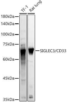
Western blot analysis of various lysates using SIGLEC3/CD33 Rabbit pAb (CAB2059) at 1:1000 dilution. Secondary antibody: HRP-conjugated Goat anti-Rabbit IgG (H+L) (AS014) at 1:10000 dilution. Lysates/proteins: 25μg per lane. Blocking buffer: 3% nonfat dry milk in TBST. Detection: ECL Basic Kit (AbGn00020). Exposure time: 3s.
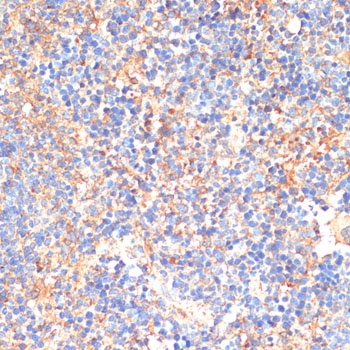
Immunohistochemistry analysis of paraffin-embedded Mouse spleen using SIGLEC3/CD33 Rabbit pAb (CAB2059) at dilution of 1:200 (40x lens). Microwave antigen retrieval performed with 0.01M PBS Buffer (pH 7.2) prior to IHC staining.
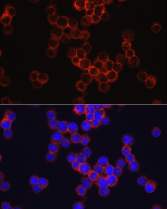
Immunofluorescence analysis of RAW264.7 cells using SIGLEC3/CD33 Rabbit pAb (CAB2059) at dilution of 1:100 (40x lens). Secondary antibody: Cy3-conjugated Goat anti-Rabbit IgG (H+L) (AS007) at 1:500 dilution. Blue: DAPI for nuclear staining.
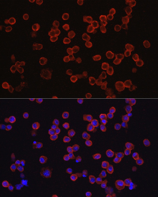
Immunofluorescence analysis of TF-1 cells using SIGLEC3/CD33 Rabbit pAb (CAB2059) at dilution of 1:100 (40x lens). Secondary antibody: Cy3-conjugated Goat anti-Rabbit IgG (H+L) (AS007) at 1:500 dilution. Blue: DAPI for nuclear staining.


")


")