The CLUH Antibody (CAB10140) is a high-quality antibody developed for reliable detection and analysis of target proteins. Enables mRNA binding activity. Involved in intracellular distribution of mitochondria. Located in cytoplasm.
This antibody is validated for use in WB, IHC-P, IF/ICC, ELISA applications and has demonstrated reactivity against Human, Mouse samples.
Product Name:
CLUH Antibody
SKU:
CAB10140
Size:
100μL, 20μL
Reactivity:
Human, Mouse
Conjugate:
Unconjugated
Immunogen:
Recombinant protein (or fragment).This information is considered to be commercially sensitive.
Tested Applications:
WBIHC-PIF/ICCELISA
Recommended Dilution:
WB
1:1000 - 1:4000
IHC-P
1:50 - 1:200
IF/ICC
1:50 - 1:200
ELISA
Recommended starting concentration is 1 μg/mL. Please optimize the concentration based on your specific assay requirements.
Synonyms:
CLU1, CLUH
Positive Sample:
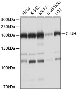
HeLa, K-562, MCF7, U-251MG, LO2
Cellular Localization:
Cytoplasm, Cytoplasmic Granule.
Calculated MW:
147kDa
Observed MW:
180kDa
Enables mRNA binding activity. Involved in intracellular distribution of mitochondria. Located in cytoplasm.
Purification Method
Affinity purification
Gene ID
23277
RRID
AB_2757667
Buffer Information
Store at -20℃. Avoid freeze / thaw cycles. Buffer: PBS containing 50% glycerol, preserved with proclin300 or sodium azide, pH 7.3.
Western blot analysis of various lysates using CLUH Rabbit pAb (CAB10140) at 1:1000 dilution. Secondary antibody: HRP-conjugated Goat anti-Rabbit IgG (H+L) (AS014) at 1:10000 dilution. Lysates/proteins: 25μg per lane. Blocking buffer: 3% nonfat dry milk in TBST. Detection: ECL Basic Kit (AbGn00020). Exposure time: 15s.
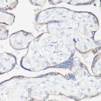
Immunohistochemistry analysis of paraffin-embedded Human placenta using CLUH Rabbit pAb (CAB10140) at dilution of 1:100 (40x lens). Microwave antigen retrieval performed with 0.01M PBS Buffer (pH 7.2) prior to IHC staining.
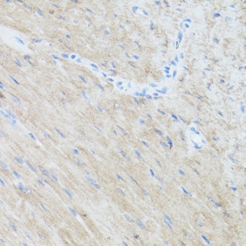
Immunohistochemistry analysis of paraffin-embedded Mouse heart using CLUH Rabbit pAb (CAB10140) at dilution of 1:100 (40x lens). Microwave antigen retrieval performed with 0.01M PBS Buffer (pH 7.2) prior to IHC staining.
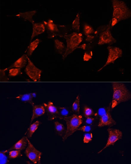
Immunofluorescence analysis of NIH-3T3 cells using CLUH Rabbit pAb (CAB10140) at dilution of 1:100 (40x lens). Secondary antibody: Cy3-conjugated Goat anti-Rabbit IgG (H+L) (AS007) at 1:500 dilution. Blue: DAPI for nuclear staining.