The COPS3 Antibody (CAB7017) is a high-quality antibody developed for reliable detection and analysis of target proteins. The protein encoded by this gene possesses kinase activity that phosphorylates regulators involved in signal transduction. It phosphorylates I kappa-Balpha, p105, and c-Jun. It acts as a docking site for complex-mediated phosphorylation. The gene is located within the Smith-Magenis syndrome region on chromosome 17. Several transcript variants encoding different isoforms have been found for this gene.
This antibody is validated for use in WB, IF/ICC, ELISA applications and has demonstrated reactivity against Human, Mouse, Rat samples.
Product Name:
COPS3 Antibody
SKU:
CAB7017
Size:
100μL, 20μL
Reactivity:
Human, Mouse, Rat
Conjugate:
Unconjugated
Immunogen:
Recombinant protein (or fragment).This information is considered to be commercially sensitive.
Tested Applications:
WBIF/ICCELISA
Recommended Dilution:
WB
1:500 - 1:2000
IF/ICC
1:50 - 1:100
ELISA
Recommended starting concentration is 1 μg/mL. Please optimize the concentration based on your specific assay requirements.
Synonyms:
CSN3, SGN3, COPS3
Positive Sample:
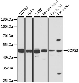
SW480, HeLa, 293T, Mouse heart, Rat heart, Rat brian
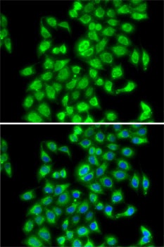
Cellular Localization:
Cytoplasm, Nucleus.
Calculated MW:
48kDa
Observed MW:
48kDa
The protein encoded by this gene possesses kinase activity that phosphorylates regulators involved in signal transduction. It phosphorylates I kappa-Balpha, p105, and c-Jun. It acts as a docking site for complex-mediated phosphorylation. The gene is located within the Smith-Magenis syndrome region on chromosome 17. Several transcript variants encoding different isoforms have been found for this gene.
Purification Method
Affinity purification
Gene ID
8533
RRID
AB_2767573
Buffer Information
Store at -20℃. Avoid freeze / thaw cycles. Buffer: PBS containing 50% glycerol, preserved with proclin300 or sodium azide, pH 7.3.
Western blot analysis of various lysates using COPS3 Rabbit pAb (CAB7017) at 1:1000 dilution. Secondary antibody: HRP-conjugated Goat anti-Rabbit IgG (H+L) (AS014) at 1:10000 dilution. Lysates/proteins: 25μg per lane. Blocking buffer: 3% nonfat dry milk in TBST. Detection: ECL Basic Kit (AbGn00020). Exposure time: 1s.
Immunofluorescence analysis of MCF7 cells using COPS3 Rabbit pAb (CAB7017). Secondary antibody: Cy3-conjugated Goat anti-Rabbit IgG (H+L) (AS007) at 1:500 dilution. Blue: DAPI for nuclear staining.