The EMR1 Antibody (CAB1256) is a high-quality antibody developed for reliable detection and analysis of target proteins. This gene encodes a protein that has a domain resembling seven transmembrane G protein-coupled hormone receptors (7TM receptors) at its C-terminus. The N-terminus of the encoded protein has six EGF-like modules, separated from the transmembrane segments by a serine/threonine-rich domain, a feature reminiscent of mucin-like, single-span, integral membrane glycoproteins with adhesive properties. Multiple alternatively spliced transcript variants encoding different isoforms have been found for this gene.
This antibody is validated for use in WB, IF/ICC, ELISA, IF-P applications and has demonstrated reactivity against Human, Mouse, Rat samples.
Product Name:
EMR1 Antibody
SKU:
CAB1256
Size:
100μL, 20μL
Reactivity:
Human, Mouse, Rat
Conjugate:
Unconjugated
Immunogen:
Recombinant protein (or fragment).This information is considered to be commercially sensitive.
Tested Applications:
WBIF/ICCELISAIF-P
Recommended Dilution:
WB
1:100 - 1:500
IF/ICC
1:50 - 1:200
IF-P
1:50 - 1:200
ELISA
Recommended starting concentration is 1 μg/mL. Please optimize the concentration based on your specific assay requirements.
Synonyms:
EMR1, TM7LN3
Positive Sample:
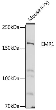
Mouse lung
Cellular Localization:
Cell Membrane, Multi-Pass Membrane Protein.
Calculated MW:
98kDa
Observed MW:
160kDa
This gene encodes a protein that has a domain resembling seven transmembrane G protein-coupled hormone receptors (7TM receptors) at its C-terminus. The N-terminus of the encoded protein has six EGF-like modules, separated from the transmembrane segments by a serine/threonine-rich domain, a feature reminiscent of mucin-like, single-span, integral membrane glycoproteins with adhesive properties. Multiple alternatively spliced transcript variants encoding different isoforms have been found for this gene.
Purification Method
Affinity purification
Gene ID
2015
RRID
AB_2759401
Buffer Information
Store at -20℃. Avoid freeze / thaw cycles. Buffer: PBS containing 50% glycerol, preserved with proclin300 or sodium azide, pH 7.3.
Western blot analysis of lysates from Mouse lung, using EMR1 Rabbit pAb (CAB1256) at 1:500 dilution. Secondary antibody: HRP-conjugated Goat anti-Rabbit IgG (H+L) (AS014) at 1:10000 dilution. Lysates/proteins: 25μg per lane. Blocking buffer: 3% nonfat dry milk in TBST. Detection: ECL Basic Kit (AbGn00020). Exposure time: 60s.
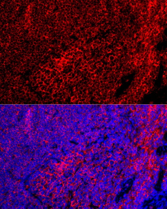
Immunofluorescence analysis of paraffin-embedded Mouse spleen using EMR1 Rabbit pAb (CAB1256) at dilution of 1:100 (40x lens). Secondary antibody: Cy3-conjugated Goat anti-Rabbit IgG (H+L) (AS007) at 1:500 dilution. Blue: DAPI for nuclear staining.
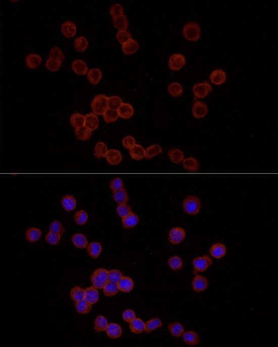
Immunofluorescence analysis of THP-1 cells using EMR1 Rabbit pAb (CAB1256) at dilution of 1:25 (40x lens). Secondary antibody: Cy3-conjugated Goat anti-Rabbit IgG (H+L) (AS007) at 1:500 dilution. Blue: DAPI for nuclear staining.