The ERK1/2 Monoclonal Antibody (CAB4782) is a high-quality antibody developed for reliable detection and analysis of target proteins. This gene encodes a member of the MAP kinase family. MAP kinases, also known as extracellular signal-regulated kinases (ERKs), act as an integration point for multiple biochemical signals, and are involved in a wide variety of cellular processes such as proliferation, differentiation, transcription regulation and development. The activation of this kinase requires its phosphorylation by upstream kinases. Upon activation, this kinase translocates to the nucleus of the stimulated cells, where it phosphorylates nuclear targets. One study also suggests that this protein acts as a transcriptional repressor independent of its kinase activity. The encoded protein has been identified as a moonlighting protein based on its ability to perform mechanistically distinct functions. Two alternatively spliced transcript variants encoding the same protein, but differing in the UTRs, have been reported for this gene. [provided by RefSeq, Jan 2014] RRID AB_2863347 Gene ID 5594 5595 Swiss Prot Synonym ERK; ERK-2; ERK2; ERT1; MAPK2; P42MAPK; PRKM1; PRKM2; p38; p40; p41; p41mapk; p42-MAPK; 5594/5595; ERK1/2
This antibody is validated for use in WB, IHC-P, IF/ICC, ELISA applications and has demonstrated reactivity against Human, Mouse, Rat samples.
Product Name:
ERK1/2 Monoclonal Antibody
SKU:
CAB4782
Size:
100μL, 20μL
Reactivity:
Human, Mouse, Rat
Clone Number:
ARC0212
Conjugate:
Unconjugated
Immunogen:
Recombinant protein (or fragment).This information is considered to be commercially sensitive.
Tested Applications:
WBIHC-PIF/ICCELISA
Recommended Dilution:
WB
1:1000 - 1:6000
IHC-P
1:4000 - 1:16000
IF
/
ICC
1:200 - 1:400
ELISA
Recommended starting concentration is 1 μg/mL. Please optimize the concentration based on your specific assay requirements.
Caveola, Cytoplasm, Cytoskeleton, Cytosol, Early Endosome, Endoplasmic Reticulum Lumen, Extracellular Region, Focal Adhesion, Golgi Apparatus, Late Endosome, Microtubule Organizing Center, Mitochondrion, Mitotic Spindle, Nucleoplasm, Nucleus, Plasma Membrane.
Calculated MW:
42 kDa/44 kDa
Observed MW:
42 kDa/44 kDa
This gene encodes a member of the MAP kinase family. MAP kinases, also known as extracellular signal-regulated kinases (ERKs), act as an integration point for multiple biochemical signals, and are involved in a wide variety of cellular processes such as proliferation, differentiation, transcription regulation and development. The activation of this kinase requires its phosphorylation by upstream kinases. Upon activation, this kinase translocates to the nucleus of the stimulated cells, where it phosphorylates nuclear targets. One study also suggests that this protein acts as a transcriptional repressor independent of its kinase activity. The encoded protein has been identified as a moonlighting protein based on its ability to perform mechanistically distinct functions. Two alternatively spliced transcript variants encoding the same protein, but differing in the UTRs, have been reported for this gene. [provided by RefSeq, Jan 2014] RRID AB_2863347 Gene ID 5594 5595 Swiss Prot Synonym ERK; ERK-2; ERK2; ERT1; MAPK2; P42MAPK; PRKM1; PRKM2; p38; p40; p41; p41mapk; p42-MAPK; 5594/5595; ERK1/2
Purification Method:
Affinity purification
Gene ID:
5594 5595
RRID:
AB_2863347
Buffer Information:
Store at -20℃. Avoid freeze / thaw cycles. Buffer: PBS containing 50% glycerol and 0.05% BSA, preserved with proclin300 or sodium azide, pH 7.3.
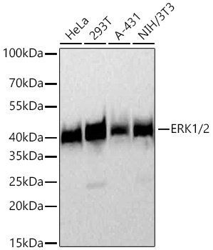
Western blot analysis of various lysates using ERK1/2 Rabbit mAb (CAB4782) at 1:4000 dilution incubated at room temperature for 1.5 hours. Secondary antibody: HRP-conjugated Goat anti-Rabbit IgG (H+L) (AS014) at 1:10000 dilution. Lysates/proteins: 25 μg per lane. Blocking buffer: 3% nonfat dry milk in TBST. Detection: ECL Basic Kit (AbGn00020). Exposure time: 45s.
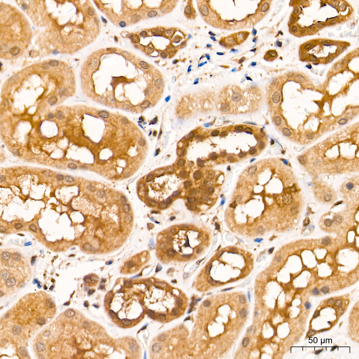
Immunohistochemistry analysis of paraffin-embedded Human kidney tissue using ERK1/2 Rabbit mAb (CAB4782) at a dilution of 1:9600 (40x lens). High pressure antigen retrieval performed with 0.01M Tris-EDTA Buffer (pH 9.0) prior to IHC staining.
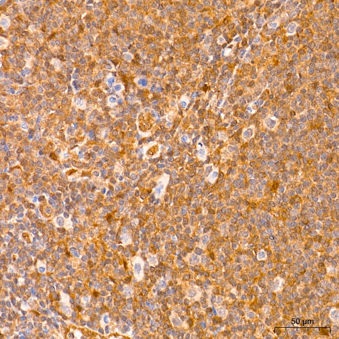
Immunohistochemistry analysis of paraffin-embedded Human tonsil tissue using ERK1/2 Rabbit mAb (CAB4782) at a dilution of 1:9600 (40x lens). High pressure antigen retrieval performed with 0.01M Tris-EDTA Buffer (pH 9.0) prior to IHC staining.
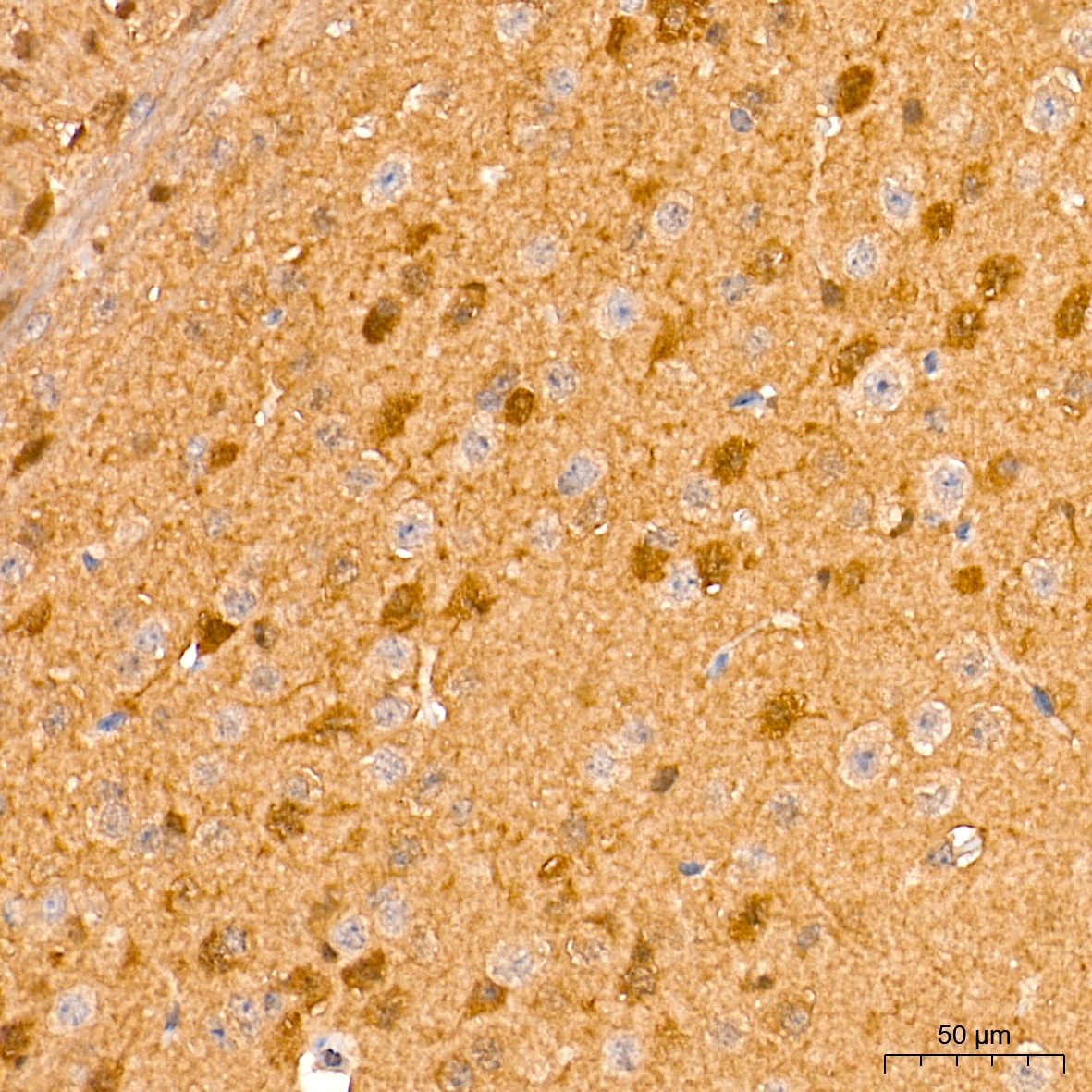
Immunohistochemistry analysis of paraffin-embedded Mouse brain tissue using ERK1/2 Rabbit mAb (CAB4782) at a dilution of 1:9600 (40x lens). High pressure antigen retrieval performed with 0.01M Tris-EDTA Buffer (pH 9.0) prior to IHC staining.