The Ferritin Light Chain Monoclonal Antibody (CAB11241) is a high-quality antibody developed for reliable detection and analysis of target proteins. This gene encodes the light subunit of the ferritin protein. Ferritin is the major intracellular iron storage protein in prokaryotes and eukaryotes. It is composed of 24 subunits of the heavy and light ferritin chains. Variation in ferritin subunit composition may affect the rates of iron uptake and release in different tissues. A major function of ferritin is the storage of iron in a soluble and nontoxic state. Defects in this light chain ferritin gene are associated with several neurodegenerative diseases and hyperferritinemia-cataract syndrome. This gene has multiple pseudogenes. RRID AB_2861532 Gene ID 2512 Swiss Prot Synonym LFTD; NBIA3; Ferritin Light Chain
This antibody is validated for use in WB, IHC-P, ELISA applications and has demonstrated reactivity against Human, Mouse, Rat samples.
Product Name:
Ferritin Light Chain Monoclonal Antibody
SKU:
CAB11241
Size:
100μL, 20μL
Reactivity:
Human, Mouse, Rat
Clone Number:
ARC57605
Conjugate:
Unconjugated
Immunogen:
Recombinant protein (or fragment).This information is considered to be commercially sensitive.
Tested Applications:
WBIHC-PELISA
Recommended Dilution:
WB
1:1000 - 1:5000
IHC-P
1:400 - 1:2000
ELISA
Recommended starting concentration is 1 μg/mL. Please optimize the concentration based on your specific assay requirements.
Synonyms:
LFTD, NBIA3, Ferritin Light Chain
Positive Sample:
Rat liver, 293T, Mouse liver, Mouse ovary, Hep G2, SH-SY5Y
This gene encodes the light subunit of the ferritin protein. Ferritin is the major intracellular iron storage protein in prokaryotes and eukaryotes. It is composed of 24 subunits of the heavy and light ferritin chains. Variation in ferritin subunit composition may affect the rates of iron uptake and release in different tissues. A major function of ferritin is the storage of iron in a soluble and nontoxic state. Defects in this light chain ferritin gene are associated with several neurodegenerative diseases and hyperferritinemia-cataract syndrome. This gene has multiple pseudogenes. RRID AB_2861532 Gene ID 2512 Swiss Prot Synonym LFTD; NBIA3; Ferritin Light Chain
Purification Method:
Affinity purification
Gene ID:
2512
RRID:
AB_2861532
Buffer Information:
Store at -20℃. Avoid freeze / thaw cycles. Buffer: PBS with 0.09% Sodium azide,0.05% BSA,50% glycerol,pH7.3.
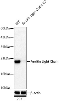
Western blot analysis of lysates from wild type (WT) and Ferritin Light Chain knockdown (KD) 293T cells using [KD Validated] Ferritin Light Chain Rabbit mAb (CAB11241) at 1:2000 dilution incubated overnight at 4℃. Secondary antibody: HRP-conjugated Goat anti-Rabbit IgG (H+L) (AS014) at 1:10000 dilution. Lysates/proteins: 25 μg per lane. Blocking buffer: 3% nonfat dry milk in TBST. Detection: ECL Basic Kit (AbGn00020). Exposure time: 60s.
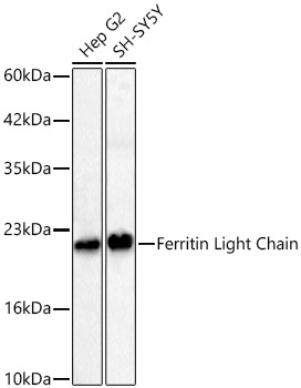
Western blot analysis of various lysates using [KD Validated] Ferritin Light Chain Rabbit mAb (CAB11241) at 1:2000 dilution incubated overnight at 4℃. Secondary antibody: HRP-conjugated Goat anti-Rabbit IgG (H+L) (AS014) at 1:10000 dilution. Lysates/proteins: 25 μg per lane. Blocking buffer: 3% nonfat dry milk in TBST. Detection: ECL Basic Kit (AbGn00020). Exposure time: 60s.
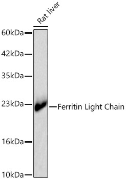
Western blot analysis of lysates from Rat liver using [KD Validated] Ferritin Light Chain Rabbit mAb (CAB11241) at 1:2000 dilution incubated overnight at 4℃. Secondary antibody: HRP-conjugated Goat anti-Rabbit IgG (H+L) (AS014) at 1:10000 dilution. Lysates/proteins: 25 μg per lane. Blocking buffer: 3% nonfat dry milk in TBST. Detection: ECL Basic Kit (AbGn00020). Exposure time: 5s.
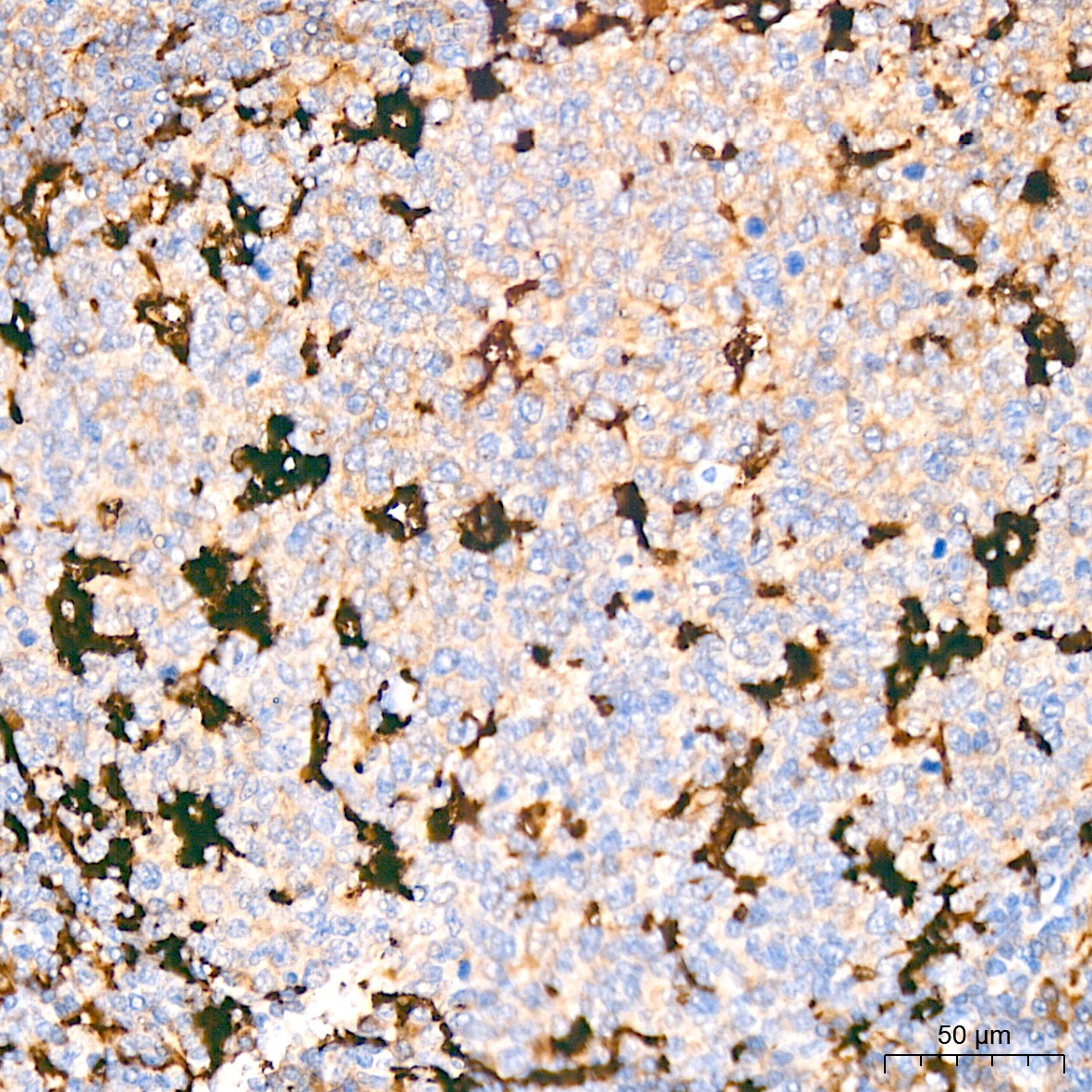
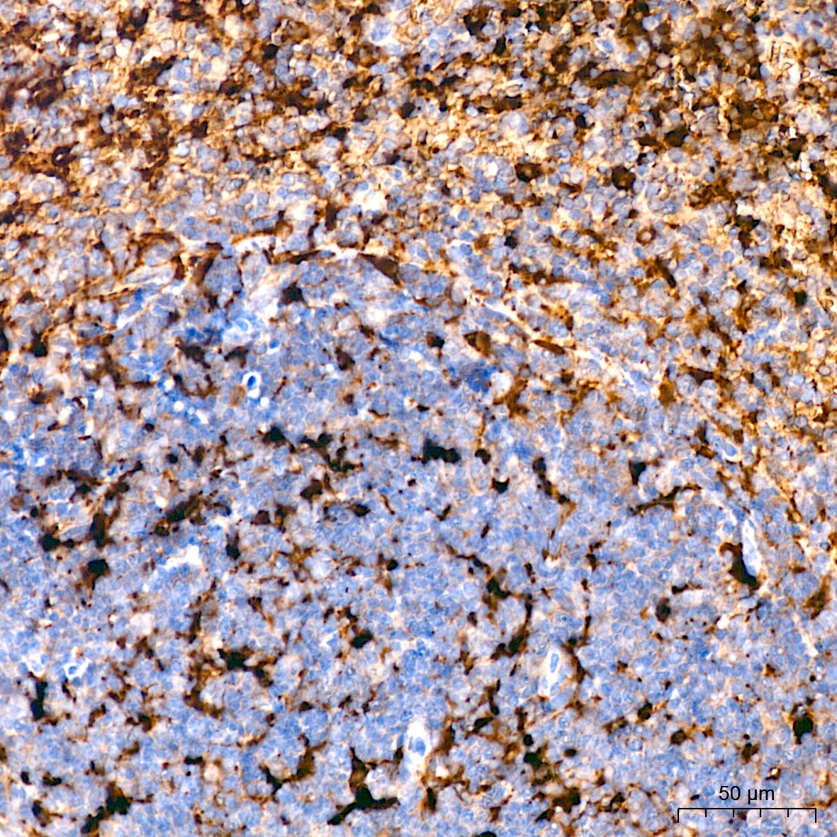
Immunohistochemistry analysis of paraffin-embedded Human tonsil tissue using [KD Validated] Ferritin Light Chain Rabbit mAb (CAB11241) at a dilution of 1:400 (40x lens). High pressure antigen retrieval performed with 0.01M Citrate buffer (pH 6.0) prior to IHC staining.
Immunohistochemistry analysis of paraffin-embedded Mouse spleen tissue using [KD Validated] Ferritin Light Chain Rabbit mAb (CAB11241) at a dilution of 1:400 (40x lens). High pressure antigen retrieval performed with 0.01M Citrate buffer (pH 6.0) prior to IHC staining.