The HER2/ErbB2 Polyclonal Antibody (CAB21768) is a high-quality antibody developed for reliable detection and analysis of target proteins. This gene encodes a member of the epidermal growth factor (EGF) receptor family of receptor tyrosine kinases. This protein has no ligand binding domain of its own and therefore cannot bind growth factors. However, it does bind tightly to other ligand-bound EGF receptor family members to form a heterodimer, stabilizing ligand binding and enhancing kinase-mediated activation of downstream signalling pathways, such as those involving mitogen-activated protein kinase and phosphatidylinositol-3 kinase. Allelic variations at amino acid positions 654 and 655 of isoform a (positions 624 and 625 of isoform b) have been reported, with the most common allele, Ile654/Ile655, shown here. Amplification and/or overexpression of this gene has been reported in numerous cancers, including breast and ovarian tumors. Alternative splicing results in several additional transcript variants, some encoding different isoforms and others that have not been fully characterized. RRID Gene ID 2064 Swiss Prot Synonym NEU; NGL; HER2; TKR1; CD340; HER-2; VSCN2; MLN 19; MLN-19; c-ERB2; c-ERB-2; HER-2/neu; p185(erbB2); B2
This antibody is validated for use in WB, ELISA applications and has demonstrated reactivity against Human samples.
Product Name:
HER2/ErbB2 Polyclonal Antibody
SKU:
CAB21768
Size:
100μL, 20μL
Reactivity:
Human
Clone Number:
-
Conjugate:
Unconjugated
Immunogen:
Synthetic peptide. This information is considered to be commercially sensitive.
Tested Applications:
WBELISA
Recommended Dilution:
WB
1:100 - 1:500
ELISA
Recommended starting concentration is 1 μg/mL. Please optimize the concentration based on your specific assay requirements.
Cell Membrane, Cytoplasm, Nucleus, Nucleus, Single-Pass Type I Membrane Protein, Perinuclear Region.
Calculated MW:
138kDa
Observed MW:
185kDa
This gene encodes a member of the epidermal growth factor (EGF) receptor family of receptor tyrosine kinases. This protein has no ligand binding domain of its own and therefore cannot bind growth factors. However, it does bind tightly to other ligand-bound EGF receptor family members to form a heterodimer, stabilizing ligand binding and enhancing kinase-mediated activation of downstream signalling pathways, such as those involving mitogen-activated protein kinase and phosphatidylinositol-3 kinase. Allelic variations at amino acid positions 654 and 655 of isoform a (positions 624 and 625 of isoform b) have been reported, with the most common allele, Ile654/Ile655, shown here. Amplification and/or overexpression of this gene has been reported in numerous cancers, including breast and ovarian tumors. Alternative splicing results in several additional transcript variants, some encoding different isoforms and others that have not been fully characterized. RRID Gene ID 2064 Swiss Prot Synonym NEU; NGL; HER2; TKR1; CD340; HER-2; VSCN2; MLN 19; MLN-19; c-ERB2; c-ERB-2; HER-2/neu; p185(erbB2); B2
Purification Method:
Affinity purification
Gene ID:
2064
RRID:
-
Buffer Information:
Store at -20℃. Avoid freeze / thaw cycles. Buffer: PBS containing 50% glycerol, preserved with proclin300 or sodium azide, pH 7.3.
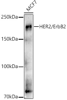
Western blot analysis of lysates from MCF7 cells, using [KO Validated] HER2/ErbB2 Rabbit pAb (CAB21768) at 1:500 dilution. Secondary antibody: HRP-conjugated Goat anti-Rabbit IgG (H+L) (AS014) at 1:10000 dilution. Lysates/proteins: 25μg per lane. Blocking buffer: 3% nonfat dry milk in TBST. Detection: ECL Basic Kit (AbGn00020). Exposure time: 180s.
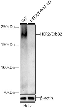
Western blot analysis of lysates from wild type(WT) and HER2/ErbB2 Rabbit pAb knockout (KO) HeLa cells, using [KO Validated] HER2/ErbB2 Rabbit pAb (CAB21768) at 1:500 dilution. Secondary antibody: HRP-conjugated Goat anti-Rabbit IgG (H+L) (AS014) at 1:10000 dilution. Lysates/proteins: 25μg per lane. Blocking buffer: 3% nonfat dry milk in TBST. Detection: ECL Basic Kit (AbGn00020). Exposure time: 180s.
 at 1:500 dilution. Secondary antibody: HRP Goat Anti-Rabbit IgG (H+L) at 1:10000 dilution. Lysates/proteins: 25μg per lane. Blocking buffer: 3% nonfat dry milk in TBST.")
 at 1:500 dilution. Secondary antibody: HRP Goat Anti-Rabbit IgG (H+L) at 1:10000 dilution. Lysates/proteins: 25μg per lane. Blocking buffer: 3% nonfat dry milk in TBST.")


")

")