The ILDR1 Polyclonal Antibody (PAC061866) is a valuable tool for researchers studying ILDR1, a protein involved in cell adhesion and signal transduction. This antibody, produced in rabbits, exhibits high specificity and sensitivity for detecting ILDR1 in human samples. Validated for use in Western blot applications, this antibody enables researchers to analyze the expression and function of ILDR1 in various cell types.ILDR1, also known as immunoglobulin-like domain containing receptor 1, is implicated in various biological processes, including cell-cell interactions and intracellular signaling pathways.
By targeting ILDR1, researchers can gain insights into its role in development, disease progression, and cellular function. This antibody is essential for investigations into the functions of ILDR1 in conditions such as cancer, immune disorders, and developmental abnormalities.Overall, the ILDR1 Polyclonal Antibody (PAC061866) is a versatile tool for researchers interested in studying the functions and mechanisms of ILDR1 in a wide range of biological contexts. Its high specificity and sensitivity make it an invaluable asset for studies in molecular biology, cell biology, and disease research.
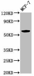
Western Blot. Positive WB detected in: MCF-7 whole cell lysate. All lanes: ILDR1 antibody at 2.6µg/ml. Secondary. Goat polyclonal to rabbit IgG at 1/50000 dilution. Predicted band size: 63, 58, 31, 24, 53, 60 kDa. Observed band size: 63 kDa.
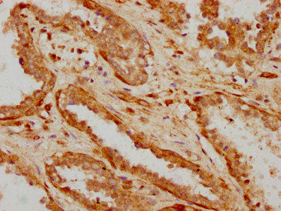
IHC image of PACO61866 diluted at 1:600 and staining in paraffin-embedded human prostate cancer performed on a Leica BondTM system. After dewaxing and hydration, antigen retrieval was mediated by high pressure in a citrate buffer (pH 6.0). Section was blocked with 10% normal goat serum 30min at RT. Then primary antibody (1% BSA) was incubated at 4°C overnight. The primary is detected by a biotinylated secondary antibody and visualized using an HRP conjugated SP system.
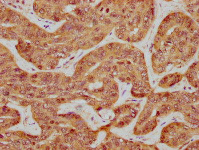
IHC image of PACO61866 diluted at 1:600 and staining in paraffin-embedded human liver cancer performed on a Leica BondTM system. After dewaxing and hydration, antigen retrieval was mediated by high pressure in a citrate buffer (pH 6.0). Section was blocked with 10% normal goat serum 30min at RT. Then primary antibody (1% BSA) was incubated at 4°C overnight. The primary is detected by a biotinylated secondary antibody and visualized using an HRP conjugated SP system.
This gene encodes a protein that contains an immunoglobulin-like domain. The encoded protein may function as a multimeric receptor at the cell surface. The expression of this gene may be a diagnostic marker for cancer progression. Alternatively spliced transcript variants encoding multiple protein isoforms have been observed for this gene. [provided by RefSeq, Dec 2010]


 ELISA Kit (HUFI03057)")
