The ULBP1 Antibody (CAB10483) is a high-quality antibody developed for reliable detection and analysis of target proteins. The protein encoded by this gene is a ligand of natural killer group 2, member D (NKG2D), an immune system-activating receptor on NK cells and T-cells. Binding of the encoded ligand to NKG2D leads to activation of several signal transduction pathways, including those of JAK2, STAT5, ERK and PI3K kinase/Akt. Also, in cytomegalovirus-infected cells, this ligand binds the UL16 glycoprotein and is prevented from activating the immune system. Three transcript variants encoding different isoforms have been found for this gene.
This antibody is validated for use in WB, IHC-P, ELISA applications and has demonstrated reactivity against Human, Mouse samples.
Product Name:
ULBP1 Antibody
SKU:
CAB10483
Size:
100μL, 20μL
Reactivity:
Human, Mouse
Conjugate:
Unconjugated
Immunogen:
Recombinant protein (or fragment).This information is considered to be commercially sensitive.
Tested Applications:
WBIHC-PELISA
Recommended Dilution:
WB
1:1000 - 1:2000
IHC-P
1:100 - 1:500
ELISA
Recommended starting concentration is 1 μg/mL. Please optimize the concentration based on your specific assay requirements.
The protein encoded by this gene is a ligand of natural killer group 2, member D (NKG2D), an immune system-activating receptor on NK cells and T-cells. Binding of the encoded ligand to NKG2D leads to activation of several signal transduction pathways, including those of JAK2, STAT5, ERK and PI3K kinase/Akt. Also, in cytomegalovirus-infected cells, this ligand binds the UL16 glycoprotein and is prevented from activating the immune system. Three transcript variants encoding different isoforms have been found for this gene.
Purification Method
Affinity purification
Gene ID
80329
RRID
AB_2758032
Buffer Information
Store at -20℃. Avoid freeze / thaw cycles. Buffer: PBS containing 50% glycerol, preserved with proclin300 or sodium azide, pH 7.3.
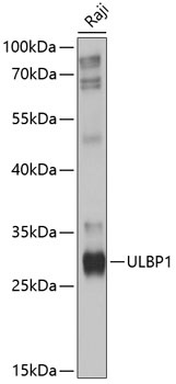
Western blot analysis of lysates from Raji cells, using ULBP1 Rabbit pAb (CAB10483) at 1:1000 dilution. Secondary antibody: HRP-conjugated Goat anti-Rabbit IgG (H+L) (AS014) at 1:10000 dilution. Lysates/proteins: 25μg per lane. Blocking buffer: 3% nonfat dry milk in TBST. Detection: ECL Basic Kit (AbGn00020). Exposure time: 10s.
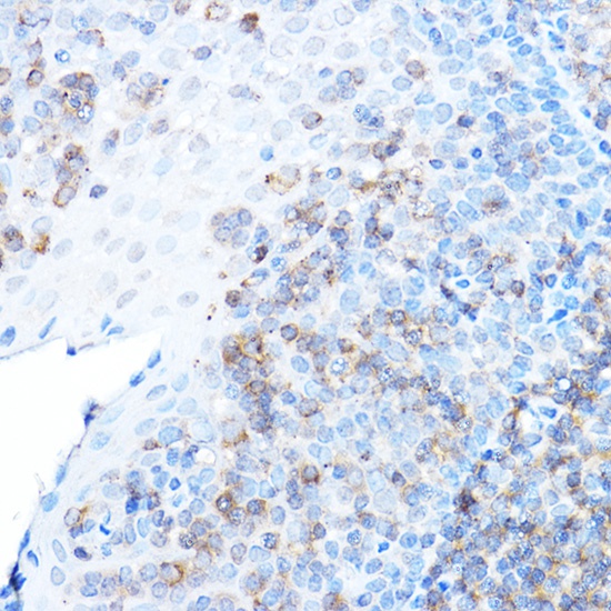
Immunohistochemistry analysis of paraffin-embedded Human tonsil using ULBP1 Rabbit pAb (CAB10483) at dilution of 1:500 (40x lens). High pressure antigen retrieval performed with 0.01M Citrate buffer (pH 6.0) prior to IHC staining.