The Phospho-CDK1-Y15 Antibody (CABP0016) is a high-quality antibody developed for reliable detection and analysis of target proteins. The protein encoded by this gene is a member of the Ser/Thr protein kinase family. This protein is a catalytic subunit of the highly conserved protein kinase complex known as M-phase promoting factor (MPF), which is essential for G1/S and G2/M phase transitions of eukaryotic cell cycle. Mitotic cyclins stably associate with this protein and function as regulatory subunits. The kinase activity of this protein is controlled by cyclin accumulation and destruction through the cell cycle. The phosphorylation and dephosphorylation of this protein also play important regulatory roles in cell cycle control. Alternatively spliced transcript variants encoding different isoforms have been found for this gene.
This antibody is validated for use in WB, IHC-P, IP, ELISA, IF-P applications and has demonstrated reactivity against Human, Mouse, Rat samples.
Product Name:
Phospho-CDK1-Y15 Antibody
SKU:
CABP0016
Size:
100μL, 20μL
Reactivity:
Human, Mouse, Rat
Conjugate:
Unconjugated
Immunogen:
Synthetic peptide. This information is considered to be commercially sensitive.
Tested Applications:
WBIHC-PIPELISAIF-P
Recommended Dilution:
WB
1:500 - 1:1000
IP
0.5μg-4μg antibody for 200μg-400μg extracts of whole cells
IF-P
1:50 - 1:200
IHC-P
1:50 - 1:200
ELISA
Recommended starting concentration is 1 μg/mL. Please optimize the concentration based on your specific assay requirements.
Synonyms:
CDC2, CDC28A, P34CDC2, Phospho-CDK1-Y15
Positive Sample:
HeLa treated with nocodazole, NIH/3T3 treated with nocodazole, C6 treated with nocodazole
Cellular Localization:
Cytoplasm, Mitochondrion, Nucleus, Centrosome, Cytoskeleton, Microtubule Organizing Center, Spindle.
Calculated MW:
34kDa
Observed MW:
34kDa
The protein encoded by this gene is a member of the Ser/Thr protein kinase family. This protein is a catalytic subunit of the highly conserved protein kinase complex known as M-phase promoting factor (MPF), which is essential for G1/S and G2/M phase transitions of eukaryotic cell cycle. Mitotic cyclins stably associate with this protein and function as regulatory subunits. The kinase activity of this protein is controlled by cyclin accumulation and destruction through the cell cycle. The phosphorylation and dephosphorylation of this protein also play important regulatory roles in cell cycle control. Alternatively spliced transcript variants encoding different isoforms have been found for this gene.
Purification Method
Affinity purification
Gene ID
983
RRID
AB_2770978
Buffer Information
Store at -20℃. Avoid freeze / thaw cycles. Buffer: PBS containing 50% glycerol, preserved with proclin300 or sodium azide, pH 7.3.
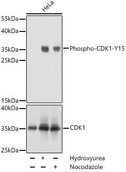
Western blot analysis of lysates from HeLa cells, using Phospho-CDK1-Y15 Rabbit pAb (A0220). HeLa cells were treated with nocodazole (50 ng/mL) at 37℃ for 20 hours or Hydroxyurea (4 mM) at 37℃ for 20 hours. Secondary antibody: HRP-conjugated Goat anti-Rabbit IgG (H+L) (AS014) at 1:10000 dilution. Lysates/proteins: 25μg per lane. Blocking buffer: 3% BSA. Detection: ECL Basic Kit (AbGn00020). Exposure time: 1s.
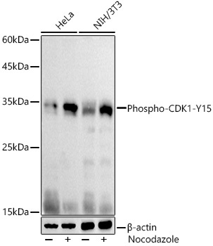
Western blot analysis of various lysates using Phospho-CDK1-Y15 Rabbit pAb (CABP0016) at 1:1000 dilution. HeLa and NIH/3T3 cells were treated with nocodazole (50 ng/ml) at 37℃ for 20 hours. Secondary antibody: HRP-conjugated Goat anti-Rabbit IgG (H+L) (AS014) at 1:10000 dilution. Lysates/proteins: 25μg per lane. Blocking buffer: 3% nonfat dry milk in TBST. Detection: ECL Basic Kit (AbGn00020). Exposure time: 10s.
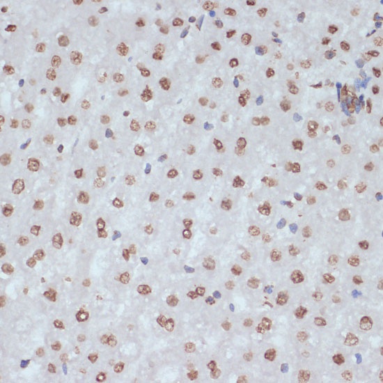
Immunohistochemistry analysis of paraffin-embedded Rat liver using Phospho-CDK1-Y15 Rabbit pAb (CABP0016) at dilution of 1:100 (40x lens). Microwave antigen retrieval performed with 0.01M Tris/EDTA Buffer (pH 9.0) prior to IHC staining.
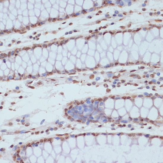
Immunohistochemistry analysis of paraffin-embedded Human colon using Phospho-CDK1-Y15 Rabbit pAb (CABP0016) at dilution of 1:100 (40x lens). Microwave antigen retrieval performed with 0.01M Tris/EDTA Buffer (pH 9.0) prior to IHC staining.
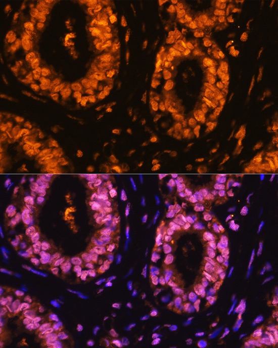
Immunofluorescence analysis of paraffin-embedded human breast cancer using Phospho-CDK1-Y15 Rabbit pAb (CABP0016) at dilution of 1:100 (40x lens). Secondary antibody: Cy3-conjugated Goat anti-Rabbit IgG (H+L) (AS007) at 1:500 dilution. Blue: DAPI for nuclear staining.
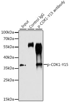
Immunoprecipitation analysis of 200 μg extracts of HT-29 cells, using 3 μg Phospho-CDK1-Y15 pAb (CABP0016). Western blot was performed from the immunoprecipitate using Phospho-CDK1-Y15 pAb (CABP0016) at a dilution of 1:1000. HT-29 cells were treated with Serum-starvation overnight at 37℃.