PIK3R6 Antibody is a premium polyclonal that offers outstanding performance and reliability for demanding research applications. Rigorously validated for ELISA, WB, IHC, IF, this antibody ensures consistent, reproducible results across multiple experimental platforms. Demonstrates excellent reactivity with Human samples, providing researchers with confidence in cross-species compatibility. Conveniently packaged in 50ug format to meet your experimental needs. For optimal performance, store at -20°C or -80°C and maintains stability for 12 months. Backed by rigorous quality control testing to ensure superior performance in your critical research applications.
Product Name:
PIK3R6 Antibody (PACO60997)
SKU:
PACO60997
Size:
50μg
Isotype:
IgG
Host Species:
Rabbit
Reactivity:
Human
Immunogen:
Recombinant Human Phosphoinositide 3-kinase regulatory subunit 6 protein (566-667AA)
Immunogen Species:
Homo sapiens (Human)
Uniprot No:
Q5UE93
Form:
Liquid
Tested Applications:
ELISAWBIHCIF
Recommended Dilution:
WB 1:500-1:5000, IHC 1:200-1:500, IF 1:50-1:200
Synonyms:
C17orf38 antibody, Chromosome 17 open reading frame 38 antibody, HsT41028 antibody, p84 PI3K adapter protein antibody, p84 PIKAP antibody, p87 phosphoinositide 3-kinase gamma (PI3Kg) adapter protein antibody, p87 PI3K adapter protein antibody, p87PIKAP antibody, Phosphatidylinositol 3-kinase, regulatory subunit 6 antibody, Phosphoinositide 3-kinase gamma adapter protein of 87 kDa antibody, Phosphoinositide 3-kinase regulatory subunit 6 antibody, PI3Kgamma adapter protein of 87 kDa antibody, PI3R6_HUMAN antibody, PIK3R6 antibody
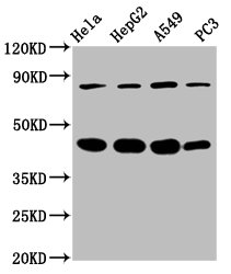
Western Blot Positive WB detected in: Hela whole cell lysate, HepG2 whole cell lysate, A549 whole cell lysate, PC-3 whole cell lysate All lanes: PIK3R6 antibody at 3.4µg/ml Secondary Goat polyclonal to rabbit IgG at 1/50000 dilution Predicted band size: 85 kDa Observed band size: 85 kDa
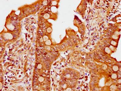
IHC image of PACO60997 diluted at 1:400 and staining in paraffin-embedded human small intestine tissue performed on a Leica BondTM system. After dewaxing and hydration, antigen retrieval was mediated by high pressure in a citrate buffer (pH 6.0). Section was blocked with 10% normal goat serum 30min at RT. Then primary antibody (1% BSA) was incubated at 4°C overnight. The primary is detected by a biotinylated secondary antibody and visualized using an HRP conjugated SP system.
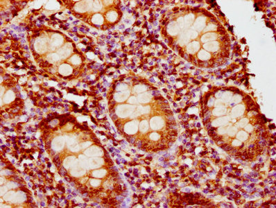
IHC image of PACO60997 diluted at 1:400 and staining in paraffin-embedded human appendix tissue performed on a Leica BondTM system. After dewaxing and hydration, antigen retrieval was mediated by high pressure in a citrate buffer (pH 6.0). Section was blocked with 10% normal goat serum 30min at RT. Then primary antibody (1% BSA) was incubated at 4°C overnight. The primary is detected by a biotinylated secondary antibody and visualized using an HRP conjugated SP system.
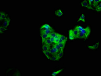
Immunofluorescence staining of HepG2 cells with PACO60997 at 1:133, counter-stained with DAPI. The cells were fixed in 4% formaldehyde, permeabilized using 0.2% Triton X-100 and blocked in 10% normal Goat Serum. The cells were then incubated with the antibody overnight at 4°C. The secondary antibody was Alexa Fluor 488-congugated AffiniPure Goat Anti-Rabbit IgG(H+L).


 ELISA Kit (HUFI03057)")


