The POLR2J Antibody (CAB1843) is a high-quality antibody developed for reliable detection and analysis of target proteins. This gene encodes a subunit of RNA polymerase II, the polymerase responsible for synthesizing messenger RNA in eukaryotes. The product of this gene exists as a heterodimer with another polymerase subunit; together they form a core subassembly unit of the polymerase. Two similar genes are located nearby on chromosome 7q22.1 and a pseudogene is found on chromosome 7p13.
This antibody is validated for use in WB, IF/ICC, ELISA applications and has demonstrated reactivity against Human, Mouse samples.
Product Name:
POLR2J Antibody
SKU:
CAB1843
Size:
100μL, 20μL
Reactivity:
Human, Mouse
Conjugate:
Unconjugated
Immunogen:
Recombinant protein (or fragment).This information is considered to be commercially sensitive.
Tested Applications:
WBIF/ICCELISA
Recommended Dilution:
WB
1:500 - 1:2000
IF/ICC
1:50 - 1:200
ELISA
Recommended starting concentration is 1 μg/mL. Please optimize the concentration based on your specific assay requirements.
Synonyms:
RPB11, RPB11A, RPB11m, hRPB14, POLR2J1, POLR2J
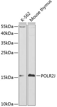
Positive Sample:
K-562, Mouse thymus
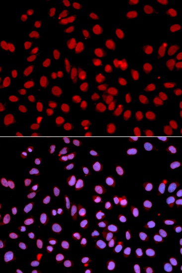
Cellular Localization:
Nucleus.
Calculated MW:
13kDa
Observed MW:
16kDa
This gene encodes a subunit of RNA polymerase II, the polymerase responsible for synthesizing messenger RNA in eukaryotes. The product of this gene exists as a heterodimer with another polymerase subunit; together they form a core subassembly unit of the polymerase. Two similar genes are located nearby on chromosome 7q22.1 and a pseudogene is found on chromosome 7p13.
Purification Method
Affinity purification
Gene ID
5439
RRID
AB_2763879
Buffer Information
Store at -20℃. Avoid freeze / thaw cycles. Buffer: PBS containing 50% glycerol, preserved with proclin300 or sodium azide, pH 7.3.
Western blot analysis of various lysates using POLR2J Rabbit pAb (CAB1843) at 1:1000 dilution. Secondary antibody: HRP-conjugated Goat anti-Rabbit IgG (H+L) (AS014) at 1:10000 dilution. Lysates/proteins: 25μg per lane. Blocking buffer: 3% nonfat dry milk in TBST.
Immunofluorescence analysis of U2OS cells using POLR2J Rabbit pAb (CAB1843). Secondary antibody: Cy3-conjugated Goat anti-Rabbit IgG (H+L) (AS007) at 1:500 dilution. Blue: DAPI for nuclear staining.