The PP1 beta Monoclonal Antibody (CAB4364) is a high-quality antibody developed for reliable detection and analysis of target proteins. The protein encoded by this gene is one of the three catalytic subunits of protein phosphatase 1 (PP1). PP1 is a serine/threonine specific protein phosphatase known to be involved in the regulation of a variety of cellular processes, such as cell division, glycogen metabolism, muscle contractility, protein synthesis, and HIV-1 viral transcription. Mouse studies suggest that PP1 functions as a suppressor of learning and memory. Two alternatively spliced transcript variants encoding distinct isoforms have been observed.
This antibody is validated for use in WB, IHC-P, IF/ICC, ELISA applications and has demonstrated reactivity against Human, Mouse, Rat samples.
Product Name:
PP1 beta Monoclonal Antibody
SKU:
CAB4364
Size:
100μL, 20μL
Reactivity:
Human, Mouse, Rat
Clone Number:
ARC0981
Conjugate:
Unconjugated
Immunogen:
Synthetic peptide. This information is considered to be commercially sensitive.
Tested Applications:
WBIHC-PIF/ICCELISA
Recommended Dilution:
WB
1:1000 - 1:6000
IHC-P
1:500 - 1:1000
IF/ICC
1:50 - 1:200
ELISA
Recommended starting concentration is 1 μg/mL. Please optimize the concentration based on your specific assay requirements.
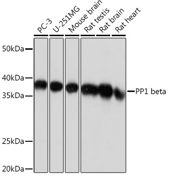
PC-3, U-251MG, Mouse brain, Rat testis, Rat brain, Rat heart
Cellular Localization:
Cytoplasm, Nucleus, Nucleolus, Nucleoplasm.
Calculated MW:
37kDa
Observed MW:
37kDa
The protein encoded by this gene is one of the three catalytic subunits of protein phosphatase 1 (PP1). PP1 is a serine/threonine specific protein phosphatase known to be involved in the regulation of a variety of cellular processes, such as cell division, glycogen metabolism, muscle contractility, protein synthesis, and HIV-1 viral transcription. Mouse studies suggest that PP1 functions as a suppressor of learning and memory. Two alternatively spliced transcript variants encoding distinct isoforms have been observed.
Purification Method
Affinity purification
Gene ID
5500
RRID
AB_2863249
Buffer Information
Store at -20℃. Avoid freeze / thaw cycles. Buffer: PBS containing 50% glycerol and 0.05% BSA, preserved with proclin300 or sodium azide, pH 7.3.
Western blot analysis of various lysates using PP1 beta Rabbit mAb (CAB4364) at 1:1000 dilution. Secondary antibody: HRP-conjugated Goat anti-Rabbit IgG (H+L) (AS014) at 1:10000 dilution. Lysates/proteins: 25μg per lane. Blocking buffer: 3% nonfat dry milk in TBST. Detection: ECL Basic Kit (AbGn00020). Exposure time: 1s.
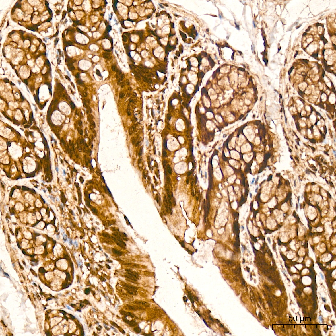
Immunohistochemistry analysis of paraffin-embedded Rat colon tissue using PP1 beta Rabbit mAb (CAB4364) at a dilution of 1:1000 (40x lens). High pressure antigen retrieval was performed with 0.01 M citrate buffer (pH 6.0) prior to IHC staining.
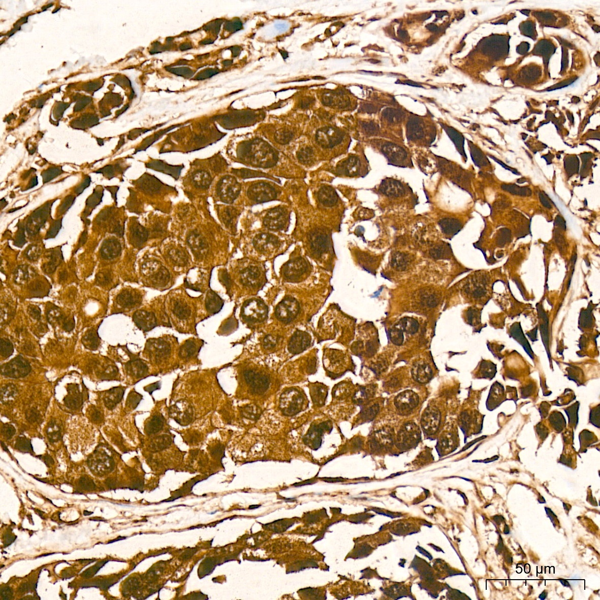
Immunohistochemistry analysis of paraffin-embedded Human breast cancer tissue using PP1 beta Rabbit mAb (CAB4364) at a dilution of 1:1000 (40x lens). High pressure antigen retrieval was performed with 0.01 M citrate buffer (pH 6.0) prior to IHC staining.
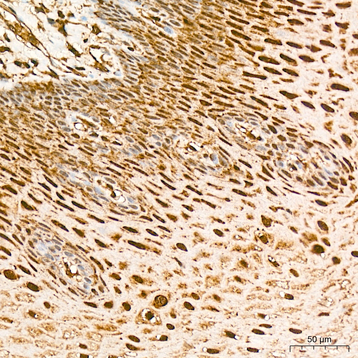
Immunohistochemistry analysis of paraffin-embedded Human esophagus tissue using PP1 beta Rabbit mAb (CAB4364) at a dilution of 1:1000 (40x lens). High pressure antigen retrieval was performed with 0.01 M citrate buffer (pH 6.0) prior to IHC staining.
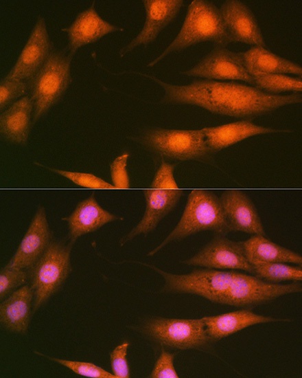
Immunofluorescence analysis of NIH-3T3 cells using PP1 beta Rabbit mAb (CAB4364) at dilution of 1:100 (40x lens). Secondary antibody: Cy3-conjugated Goat anti-Rabbit IgG (H+L) (AS007) at 1:500 dilution. Blue: DAPI for nuclear staining.
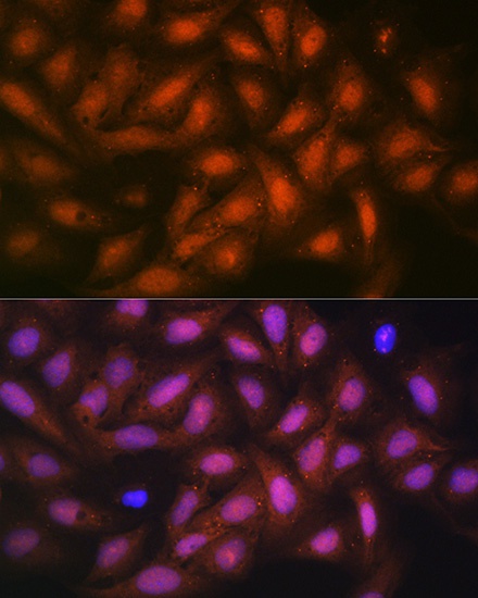
Immunofluorescence analysis of U-2 OS cells using PP1 beta Rabbit mAb (CAB4364) at dilution of 1:100 (40x lens). Secondary antibody: Cy3-conjugated Goat anti-Rabbit IgG (H+L) (AS007) at 1:500 dilution. Blue: DAPI for nuclear staining.