The PTGFR Antibody (CAB15088) is a high-quality antibody developed for reliable detection and analysis of target proteins. The protein encoded by this gene is member of the G-protein coupled receptor family. This protein is a receptor for prostaglandin F2-alpha (PGF2-alpha), which is known to be a potent luteolytic agent, and may also be involved in modulating intraocular pressure and smooth muscle contraction in uterus. Knockout studies in mice suggest that the interaction of PGF2-alpha with this receptor may initiate parturition in ovarian luteal cells and thus induce luteolysis. Two transcript variants encoding different isoforms have been found for this gene.
This antibody is validated for use in WB, ELISA applications and has demonstrated reactivity against Mouse, Rat samples.
Product Name:
PTGFR Antibody
SKU:
CAB15088
Size:
100μL, 20μL
Reactivity:
Mouse, Rat
Conjugate:
Unconjugated
Immunogen:
Synthetic peptide. This information is considered to be commercially sensitive.
Tested Applications:
WBELISA
Recommended Dilution:
WB
1:500 - 1:2000
ELISA
Recommended starting concentration is 1 μg/mL. Please optimize the concentration based on your specific assay requirements.
Synonyms:
FP, PTGFR
Positive Sample:
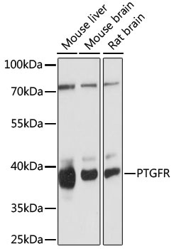
mouse liver, mouse brain, rat brain
Cellular Localization:
Cell Membrane, Multi-Pass Membrane Protein.
Calculated MW:
40kDa
Observed MW:
40kDa
The protein encoded by this gene is member of the G-protein coupled receptor family. This protein is a receptor for prostaglandin F2-alpha (PGF2-alpha), which is known to be a potent luteolytic agent, and may also be involved in modulating intraocular pressure and smooth muscle contraction in uterus. Knockout studies in mice suggest that the interaction of PGF2-alpha with this receptor may initiate parturition in ovarian luteal cells and thus induce luteolysis. Two transcript variants encoding different isoforms have been found for this gene.
Purification Method
Affinity purification
Gene ID
5737
RRID
AB_2761971
Buffer Information
Store at -20℃. Avoid freeze / thaw cycles. Buffer: PBS with 0.01% thimerosal,50% glycerol,pH7.3.
Western blot analysis of various lysates using PTGFR Rabbit pAb (CAB15088) at 1:1000 dilution. Secondary antibody: HRP-conjugated Goat anti-Rabbit IgG (H+L) (AS014) at 1:10000 dilution. Lysates/proteins: 25μg per lane. Blocking buffer: 3% nonfat dry milk in TBST. Detection: ECL Basic Kit (AbGn00020). Exposure time: 90s.