The RCN1 Polyclonal Antibody (CAB21590) is a high-quality antibody developed for reliable detection and analysis of target proteins. Reticulocalbin 1 is a calcium-binding protein located in the lumen of the ER. The protein contains six conserved regions with similarity to a high affinity Ca(+2)-binding motif, the EF-hand. High conservation of amino acid residues outside of these motifs, in comparison to mouse reticulocalbin, is consistent with a possible biochemical function besides that of calcium binding. In human endothelial and prostate cancer cell lines this protein localizes to the plasma membrane.
This antibody is validated for use in WB, IF/ICC, ELISA applications and has demonstrated reactivity against Human, Mouse samples.
Product Name:
RCN1 Polyclonal Antibody
SKU:
CAB21590
Size:
100μL, 20μL
Reactivity:
Human, Mouse
Conjugate:
Unconjugated
Immunogen:
Recombinant protein (or fragment).This information is considered to be commercially sensitive.
Tested Applications:
WBIF/ICCELISA
Recommended Dilution:
WB
1:500 - 1:2000
IF/ICC
1:50 - 1:200
ELISA
Recommended starting concentration is 1 μg/mL. Please optimize the concentration based on your specific assay requirements.
Synonyms:
RCN, RCAL, PIG20, HEL-S-84, RCN1
Positive Sample:
A549, SK-OV-3, L-O2, HeLa
Cellular Localization:
Endoplasmic Reticulum Lumen.
Calculated MW:
39kDa
Observed MW:
Refer to figures
Reticulocalbin 1 is a calcium-binding protein located in the lumen of the ER. The protein contains six conserved regions with similarity to a high affinity Ca(+2)-binding motif, the EF-hand. High conservation of amino acid residues outside of these motifs, in comparison to mouse reticulocalbin, is consistent with a possible biochemical function besides that of calcium binding. In human endothelial and prostate cancer cell lines this protein localizes to the plasma membrane.
Purification Method
Affinity purification
Gene ID
5954
Buffer Information
Store at -20℃. Avoid freeze / thaw cycles. Buffer: PBS containing 50% glycerol, preserved with proclin300 or sodium azide, pH 7.3.
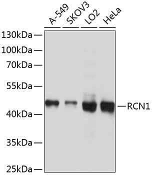
Western blot analysis of various lysates using RCN1 Rabbit pAb (CAB21590) at 1:1000 dilution. Secondary antibody: HRP-conjugated Goat anti-Rabbit IgG (H+L) (AS014) at 1:10000 dilution. Lysates/proteins: 25μg per lane. Blocking buffer: 3% nonfat dry milk in TBST. Detection: ECL Basic Kit (AbGn00020). Exposure time: 1s.
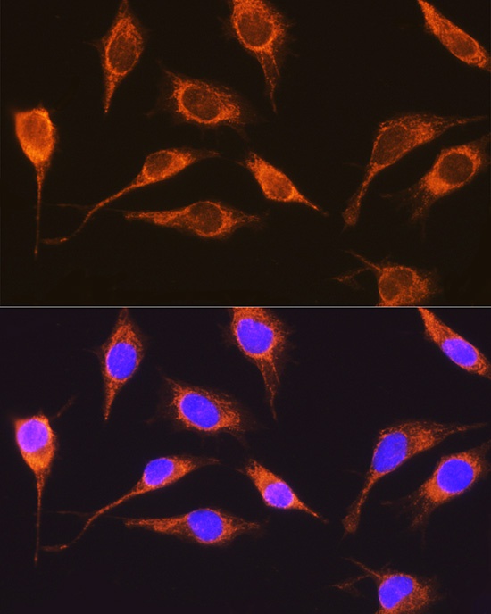
Immunofluorescence analysis of L929 cells using RCN1 Rabbit pAb (CAB21590) at dilution of 1:100 (40x lens). Secondary antibody: Cy3-conjugated Goat anti-Rabbit IgG (H+L) (AS007) at 1:500 dilution. Blue: DAPI for nuclear staining.
 at 1:1000 dilution. Secondary antibody: HRP Goat Anti-Rabbit IgG (H+L) at 1:10000 dilution. Lysates/proteins: 25μg per lane. Blocking buffer: 3% nonfat dry milk in TBST.")
 at 1:1000 dilution. Secondary antibody: HRP Goat Anti-Rabbit IgG (H+L) at 1:10000 dilution. Lysates/proteins: 25μg per lane. Blocking buffer: 3% nonfat dry milk in TBST.")


 ELISA Kit (HUES03544)")
 at 1:10000 dilution. Lysates/proteins: 25ug per lane. Blocking buffer: 3% nonfat dry milk in TBST. Detection: ECL Enhanced Kit. Exposure time: 300s.")

")