")
Description
Recombinant Human DC-SIGN/CD209 Protein
The Recombinant Human DC-SIGN/CD209 Protein is a biologically active recombinant protein that plays a significant role in various cellular processes and signaling pathways in human biology. This protein is widely employed in immunological research, cell biology studies, protein-protein interaction analyses, and therapeutic development, providing researchers with a reliable tool for investigating DC-SIGN/CD209 function and its implications in health and disease.
This product (SKU: RPCB1439) is produced using HEK293 cells and features a C-His tag for convenient detection and purification. The protein exhibits a calculated molecular weight of 40.28 kDa with an observed molecular weight of 40-50 kDa under denaturing conditions, achieving ≥ 95 % as determined by SDS-PAGE.. Functional bioactivity has been validated through rigorous quality control assays, confirming its suitability for demanding research applications.
Key Features
| High Purity by Affinity Chromatography | |
| Mammalian & Bacterial Expression Systems | |
| High lot-to-lot consistency via strict QC |
| Product Name: | Recombinant Human DC-SIGN/CD209 Protein |
| SKU: | RPCB1439 |
| Size: | 10 μg , 20 μg , 50 μg , 100 μg |
| Reactivity: | Human |
| Synonyms: | CD209, CDSIGN, CLEC4L, DC-SIGN, DC-SIGN1, CLEC4L, DC-SIGN, DC-SIGN1 |
| Tag: | C-His |
| Expression Host: | HEK293 cells |
| Calculated MW: | 40.28 kDa |
| Observed MW: | 40-50 kDa |
| Gene ID: | 30835 |
| Protein Description: | High quality, high purity and low endotoxin recombinant Recombinant Human DC-SIGN/CD209 Protein (RPCB1439), tested reactivity in HEK293 cells and has been validated in SDS-PAGE.100% guaranteed. |
| Endotoxin: | < 0.1 EU/μg of the protein by LAL method. |
| Purity: | ≥ 95 % as determined by SDS-PAGE. |
| Formulation: | Lyophilized from a 0.22 μm filtered solution of PBS, pH 7.4. |
| Bio-Activity: | Measured by the ability of the immobilized protein to support the adhesion of ICAM-3 expressing CHO Chinese hamster ovary cells. When 5 x 10⁴ cells/well are added to Recombinant Human DC_x001E_SIGN/CD209 Protein coated plates (5 μg/mL with 100 μL/well), approximately 10-20% of added cells will adhere after 1 hour at 37℃. |
| Reconstitution: | Centrifuge the vial before opening. Reconstitute to a concentration of 0.1-0.5 mg/mL in sterile distilled water. Avoid vortex or vigorously pipetting the protein. For long term storage, it is recommended to add a carrier protein or stablizer (e.g. 0.1% BSA, 5% HSA, 10% FBS or 5% Trehalose), and aliquot the reconstituted protein solution to minimize free-thaw cycles. |
| Storage: | Store at -20℃.Store the lyophilized protein at -20℃ to -80 ℃ up to 1 year from the date of receipt. After reconstitution, the protein solution is stable at -20℃ for 3 months, at 2-8℃ for up to 1 week. |
Dendritic cell (DC)-specific intercellular adhesion molecule 3 (ICAM-3) grabbing nonintegrin (DC-SIGN), also known as CD209, is a type II transmembrane protein on DCs with a C-type lectin extracellular domain, is capable of binding ICAM-3 on resting T cells in the secondary lymphoid organs, providing the initial contact between these cells during the establishment of cell-mediated immunity. It is not only a pattern recognition receptor but implicated in immunoregulation of DCs. It has an important role in mediating DC adhesion, migration, inflammation, activating primary T cell, triggering immune response and participating in immune escape of pathogens and tumors. DC-SIGN also mediates the capture and internalization of viral, bacterial, and fungal pathogens by dendritic cells, such as HIV-1, Ebola virus, cytomegalovirus, Dengue virus, and hepatitis C virus. DC-SIGN is unique in that it regulates adhesion processes, such as DC trafficking and T-cell synapse formation, as well as antigen capture. Moreover, even though several C-type lectins have been shown to bind HIV-1, DC-SIGN does not only capture HIV-1 but also protects it in early endosomes allowing HIV-1 transport by DC to lymphoid tissues, where it enhances trans infection of T cells.
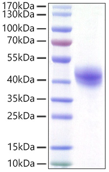 | Recombinant Human DC-SIGN/CD209 Protein was determined by SDS-PAGE under reducing conditions with Coomassie Blue. |
")
 (RPES5123)")
")

")
 ELISA Kit (HUEB1985)")