")
Description
Recombinant Mouse TNFSF15/TL1A Protein
The Recombinant Mouse TNFSF15/TL1A Protein is a high-quality recombinant protein designed for murine biological research applications. This protein serves as an essential reagent in mouse model studies, comparative immunology research, and preclinical therapeutic evaluations, enabling scientists to investigate TNFSF15/TL1A biology and its relevance to human disease mechanisms through translational research approaches.
This product (SKU: RPCB0013) is produced using advanced expression systems and features a NO-Tag tag for convenient detection and purification. The protein exhibits a calculated molecular weight of 19.90 kDa with an observed molecular weight of 20-25 kDa under denaturing conditions, achieving ≥ 95 % as determined by SDS-PAGE.. Functional bioactivity has been validated through rigorous quality control assays, confirming its suitability for demanding research applications.
Key Features
| High Purity by Affinity Chromatography | |
| Mammalian & Bacterial Expression Systems | |
| High lot-to-lot consistency via strict QC |
| Product Name: | Recombinant Mouse TNFSF15/TL1A Protein |
| SKU: | RPCB0013 |
| Size: | 10 μg |
| Reactivity: | Mouse |
| Synonyms: | Tnfsf15, Tl1, Vegi, Tumor necrosis factor ligand superfamily member 15, TNF ligand-related molecule 1, Vascular endothelial cell growth inhibitor |
| Tag: | NO-Tag |
| Calculated MW: | 19.90 kDa |
| Observed MW: | 20-25 kDa |
| Gene ID: | 326623 |
| Protein Description: | High quality, high purity and low endotoxin recombinant Recombinant Mouse TNFSF15/TL1A Protein (RPCB0013), tested reactivity in E. coli and has been validated in SDS-PAGE.100% guaranteed. |
| Endotoxin: | < 1 EU/μg of the protein by LAL method. |
| Purity: | ≥ 95 % as determined by SDS-PAGE. |
| Formulation: | Lyophilized from 0.22 μm filtered solution in 20mM PB,300mM NaCl (pH 7.0). Normally 8% trehalose is added as protectant before lyophilization. |
| Bio-Activity: | Measured by its ability to induce apoptosis of TF‑1 human erythroleukemic cells. The ED 50 for this effect is 15.61-62.44 ng/mL, corresponding to a specific activity of 1.60×10 4 ~6.41×10 4 units/mg. |
| Reconstitution: | Centrifuge the vial before opening. Reconstitute to a concentration of 0.1-0.5 mg/mL in sterile distilled water. Avoid vortex or vigorously pipetting the protein. For long term storage, it is recommended to add a carrier protein or stablizer (e.g. 0.1% BSA, 5% HSA, 10% FBS or 5% Trehalose), and aliquot the reconstituted protein solution to minimize free-thaw cycles. |
| Storage: | Store at -20℃.Store the lyophilized protein at -20℃ to -80 ℃ up to 1 year from the date of receipt. After reconstitution, the protein solution is stable at -20℃ for 3 months, at 2-8℃ for up to 1 week. |
TL1A is a type II transmembrane protein belonging to the TNF superfamily and has been designated TNF superfamily member 15 (TNFSF15). Mouse TL1A is a 252 amino acid residues (aa) protein consisting of a 35 aa N-terminal cytoplasmic domain, a 24 aa transmembrane region and 193 aa C-terminal extracellular domain. TL1A is expressed predominantly in endothelial cells and its expression is stimulated by TNF-a and IL-1a. Non-endothelial cells of the gut mucosa, including lamina propria lymphocytes and tissue macrophages, also express TL1A and at higher levels in chronic inflammatory bowel disorders. TL1A binds with high affinity to death receptor 3 (DR3), which is now designated TNF receptor superfamily member 25 (TNFRSF25). DR3 was formerly designated TNFRSF12 when it was thought to be the receptor for TWEAK/TNFSF12. Depending on the cell type, DR3-TL1A interactions have different effects. Ligation of DR3 on activated T cells by TL1A provides a costimulatory signal to increase IL-2 responsiveness and the secretion of proinflamatory cytokines. The promotion of survival of activated T cells by TL1a results from the activation of the transcription factor NF-kappa-B. In a tumor erythroleukemic cell line, TF-1, DR3-TL1A signaling increases production of the NF-kB-dependent antiapoptotic protein c-IAP2, promoting survival in these cells. In HUVEC cells, which express both DR3 and TL1A, ligation of DR3 by TL1A regulates cell apoptosis. These effects of TL1A are blocked by the secreted, soluble decoy receptor 3 (DcR3), also known as TR6 and TNFRSF6B, which compete with DR3 for binding to TL1A. Consistent with the observed in vitro activities, TL1A promotes ex vivo splenocyte expansion and enhances in vivo graft-versus-host-response. The level of TL1A in cells of gut mucosa, in patients with bowel inflammatory disorders, correlates with the severity of inflammation, and TL1A may play a role in a Th1-mediate pathological conditions such as Crohn’s disease.
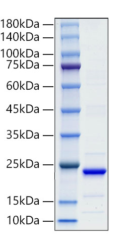 | Recombinant Mouse TNFSF15/TL1A Protein was determined by SDS-PAGE under reducing (R). |
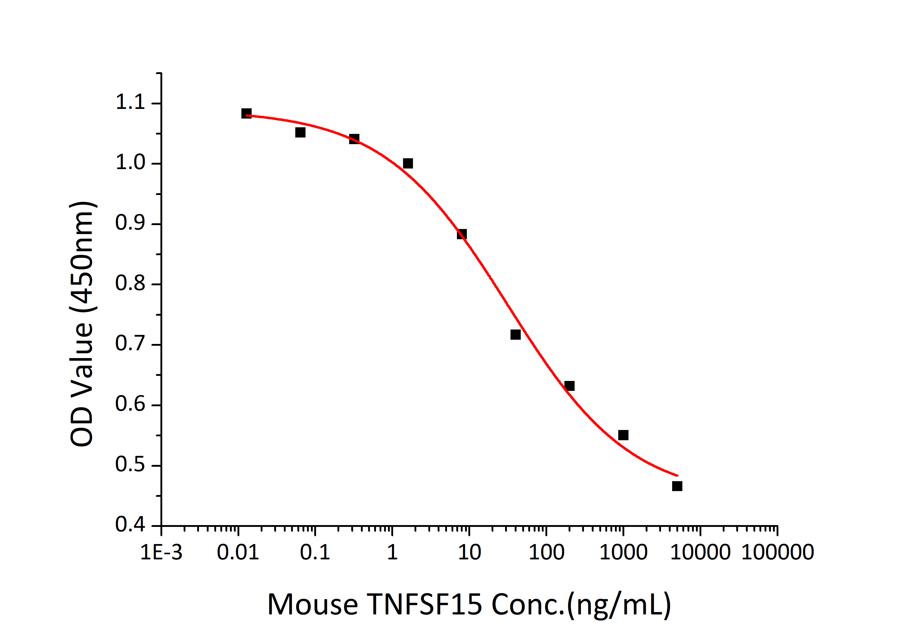 | Recombinant Mouse TNFSF15/TL1A Protein induce apoptosis of TF‑1 human erythroleukemic cells. The ED50 for this effect is 15.61-62.44 ng/mL, corresponding to a specific activity of 1.60×10⁴~6.41×10⁴ units/mg. |
")


")
")
")