The RPS10 Antibody (CAB6056) is a high-quality antibody developed for reliable detection and analysis of target proteins. Ribosomes, the organelles that catalyze protein synthesis, consist of a small 40S subunit and a large 60S subunit. Together these subunits are composed of 4 RNA species and approximately 80 structurally distinct proteins. This gene encodes a ribosomal protein that is a component of the 40S subunit. The protein belongs to the S10E family of ribosomal proteins. It is located in the cytoplasm. Variable expression of this gene in colorectal cancers compared to adjacent normal tissues has been observed, although no correlation between the level of expression and the severity of the disease has been found. As is typical for genes encoding ribosomal proteins, there are multiple processed pseudogenes of this gene dispersed through the genome. Alternate splicing results in multiple transcript variants that encode the same protein. Naturally occurring read-through transcription occurs between this locus and the neighboring locus NUDT3 (nudix (nucleoside diphosphate linked moiety X)-type motif 3). RRID AB_2766730 Gene ID 6204 Swiss Prot Synonym S10; DBA9; eS10; RPS10
This antibody is validated for use in WB, IHC-P, IF/ICC, ELISA applications and has demonstrated reactivity against Human, Mouse, Rat samples.
Product Name:
RPS10 Antibody
SKU:
CAB6056
Size:
100μL, 20μL
Reactivity:
Human, Mouse, Rat
Clone Number:
-
Conjugate:
Unconjugated
Immunogen:
Recombinant protein (or fragment).This information is considered to be commercially sensitive.
Tested Applications:
WBIHC-PIF/ICCELISA
Recommended Dilution:
WB
1:500 - 1:1000
IHC-P
1:20 - 1:200
IF
/
ICC
1:20 - 1:100
ELISA
Recommended starting concentration is 1 μg/mL. Please optimize the concentration based on your specific assay requirements.
Synonyms:
S10, DBA9, eS10, RPS10
Positive Sample:
HeLa, K-562, Mouse liver, Mouse thymus, Rat thymus
Cellular Localization:
Cytoplasm, Nucleus, Nucleolus.
Calculated MW:
19kDa
Observed MW:
19kDa
Ribosomes, the organelles that catalyze protein synthesis, consist of a small 40S subunit and a large 60S subunit. Together these subunits are composed of 4 RNA species and approximately 80 structurally distinct proteins. This gene encodes a ribosomal protein that is a component of the 40S subunit. The protein belongs to the S10E family of ribosomal proteins. It is located in the cytoplasm. Variable expression of this gene in colorectal cancers compared to adjacent normal tissues has been observed, although no correlation between the level of expression and the severity of the disease has been found. As is typical for genes encoding ribosomal proteins, there are multiple processed pseudogenes of this gene dispersed through the genome. Alternate splicing results in multiple transcript variants that encode the same protein. Naturally occurring read-through transcription occurs between this locus and the neighboring locus NUDT3 (nudix (nucleoside diphosphate linked moiety X)-type motif 3). RRID AB_2766730 Gene ID 6204 Swiss Prot Synonym S10; DBA9; eS10; RPS10
Purification Method:
Affinity purification
Gene ID:
6204
RRID:
AB_2766730
Buffer Information:
Store at -20℃. Avoid freeze / thaw cycles. Buffer: PBS containing 50% glycerol, preserved with proclin300 or sodium azide, pH 7.3.
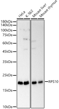
Western blot analysis of various lysates, using RPS10 Rabbit pAb (CAB6056) at 1:1000 dilution. Secondary antibody: HRP-conjugated Goat anti-Rabbit IgG (H+L) (AS014) at 1:10000 dilution. Lysates/proteins: 25μg per lane. Blocking buffer: 3% nonfat dry milk in TBST. Detection: ECL Basic Kit (AbGn00020). Exposure time: 20s.
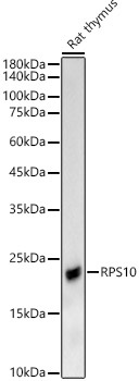
Western blot analysis of lysates from Rat thymus, using RPS10 Rabbit pAb (CAB6056) at 1:1000 dilution. Secondary antibody: HRP-conjugated Goat anti-Rabbit IgG (H+L) (AS014) at 1:10000 dilution. Lysates/proteins: 25μg per lane. Blocking buffer: 3% nonfat dry milk in TBST. Detection: ECL Basic Kit (AbGn00020). Exposure time: 90s.
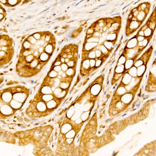
Immunohistochemistry analysis of paraffin-embedded Rat colon tissue using RPS10 Rabbit pAb (CAB6056) at a dilution of 1:100 (40x lens). High pressure antigen retrieval was performed with 0.01 M citrate buffer (pH 6.0) prior to IHC staining.
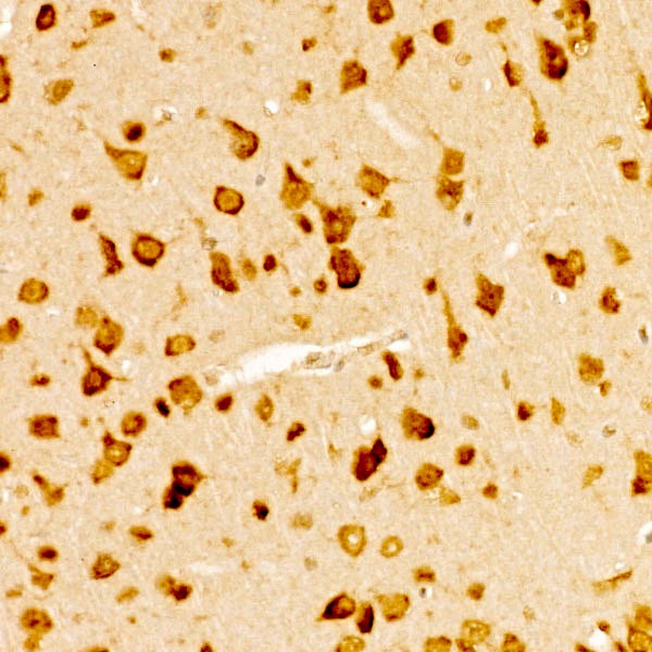
Immunohistochemistry analysis of paraffin-embedded Mouse brain tissue using RPS10 Rabbit pAb (CAB6056) at a dilution of 1:100 (40x lens). High pressure antigen retrieval was performed with 0.01 M citrate buffer (pH 6.0) prior to IHC staining.