SBK2 Antibody is a premium polyclonal that offers outstanding performance and reliability for demanding research applications. Rigorously validated for ELISA, IHC, IF, this antibody ensures consistent, reproducible results across multiple experimental platforms. Demonstrates excellent reactivity with Human samples, providing researchers with confidence in cross-species compatibility. Conveniently packaged in 50ul format to meet your experimental needs. For optimal performance, store at -20°C or -80°C and maintains stability for 12 months. Backed by rigorous quality control testing to ensure superior performance in your critical research applications.
Product Name:
SBK2 Antibody (PACO62371)
SKU:
PACO62371
Size:
50μl
Isotype:
IgG
Host Species:
Rabbit
Reactivity:
Human
Immunogen:
Recombinant Human Serine/threonine-protein kinase SBK2 protein (62-330AA)
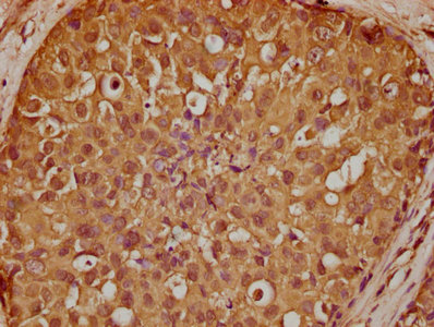
IHC image of PACO62371 diluted at 1:100 and staining in paraffin-embedded human kidney tissue performed on a Leica BondTM system. After dewaxing and hydration, antigen retrieval was mediated by high pressure in a citrate buffer (pH 6.0). Section was blocked with 10% normal goat serum 30min at RT. Then primary antibody (1% BSA) was incubated at 4°C overnight. The primary is detected by a biotinylated secondary antibody and visualized using an HRP conjugated SP system.
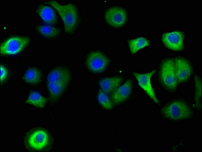
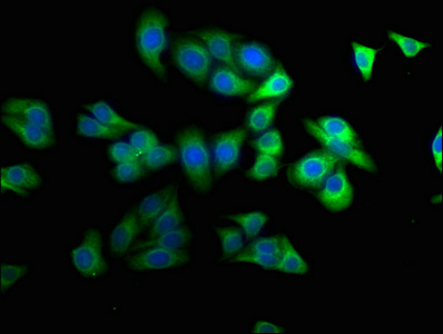
Immunofluorescence staining of A549 cells with PACO62371 at 1:50, counter-stained with DAPI. The cells were fixed in 4% formaldehyde, permeabilized using 0.2% Triton X-100 and blocked in 10% normal Goat Serum. The cells were then incubated with the antibody overnight at 4°C. The secondary antibody was Alexa Fluor 488-congugated AffiniPure Goat Anti-Rabbit IgG(H+L).
IHC image of PACO62371 diluted at 1:200 and staining in paraffin-embedded human breast cancer performed on a Leica BondTM system. After dewaxing and hydration, antigen retrieval was mediated by high pressure in a citrate buffer (pH 6.0). Section was blocked with 10% normal goat serum 30min at RT. Then primary antibody (1% BSA) was incubated at 4°C overnight. The primary is detected by a biotinylated secondary antibody and visualized using an HRP conjugated SP system.
Immunofluorescence staining of HepG2 cells with PACO62371 at 1:100, counter-stained with DAPI. The cells were fixed in 4% formaldehyde, permeabilized using 0.2% Triton X-100 and blocked in 10% normal Goat Serum. The cells were then incubated with the antibody overnight at 4°C. The secondary antibody was Alexa Fluor 488-congugated AffiniPure Goat Anti-Rabbit IgG(H+L).


 ELISA Kit (HUFI03057)")

